SlideShare uma empresa Scribd logo
1 de 14
Baixar para ler offline
Appropriate Use of Anti-Aβ Monoclonal Antibodies in
Clinical Practice: Baseline Radiographic Exclusion Criteria1,2
Full abbreviations, accreditation, and disclosure information available at PeerView.com/MCV40
1. Cummings J et al. J Prev Alzheimers Dis. 2023;10:362-377. 2. Cummings J et al. J Prev Alzheimers Dis. 2022;2:221-230.
• >4 microhemorrhages (defined as ≤10 mm at the greatest diameter)
• A single macrohemorrhage >10 mm at greatest diameter
• An area of superficial siderosis
• Evidence of vasogenic edema
• >2 lacunar infarcts or stroke involving a major vascular territory
• Severe subcortical hyperintensities consistent with a Fazekas score of 3
• Evidence of ABRA
• Evidence of CAA-ri
• Any other major intracranial pathology that may cause cognitive impairment (eg, cerebral contusion,
encephalomalacia, brain aneurysms or other vascular malformations, CNS infection, and brain tumors
other than meningioma or arachnoid cysts)
• MRI evidence of a non-AD dementia
Baseline MRI-Based Exclusion Criteria
Detecting, Monitoring, and Managing Amyloid-Related
Imaging Abnormalities (ARIA)
Full abbreviations, accreditation, and disclosure information available at PeerView.com/MCV40
3T scanner (recommended)
1.5T scanner (minimal)
High field strength scanners have greater sensitivity but limited availability
The use of 1.5T scanner is endorsed as a minimum standard
Slice thickness: ≤5 mm Thinner slices increase resolution, but decrease signal-to-noise ratio
TE: ≥20 ms Longer TE increases sensitivity to detection
2D T2* GRE or SWI
(for ARIA-H)
MRI sequences used to improve the detection, visualization, and monitoring of
microhemorrhages and superficial siderosis (ARIA-H)
T2-FLAIR (for ARIA-E) MRI sequences used to detect and monitor brain edema or sulcal effusion (ARIA-E)
Diffusion-weighted imaging
An MRI method used to differentiate between ARIA and cytotoxic processes (eg, infarcts)
Recommended for differential diagnosis
Visit the American Society for Neuroradiology (ASNR) website to access ARIA resources,
including links to standardized protocols for 3T and 1.5T scanners via this QR code or URL:
https://www.asnr.org/education-resources/alzheimers-webinar-series
MRI Acquisition Protocols to Detect and Monitor ARIA1-3
Detecting, Monitoring, and Managing Amyloid-Related
Imaging Abnormalities (ARIA)
Full abbreviations, accreditation, and disclosure information available at PeerView.com/MCV40
ARIA Type
ARIA-E
ARIA-H
microhemorrhage
ARIA-H superficial
siderosis
FLAIR hyperintensity
confined to sulcus and/or
cortex/subcortical white
matter in one location <5 cm
Mild Moderate
Radiographic Severity
Severe
≤4 new incident
microhemorrhages
One focal area
of superficial siderosis
FLAIR hyperintensity 5-10 cm,
or more than one site
of involvement, each
measuring <10 cm
5-9 new incident
microhemorrhages
Two focal areas
of superficial siderosis
FLAIR hyperintensity measures
>10 cm, often with significant
subcortical white matter
and/or sulcal involvement;
≥1 separate sites of
involvement might be noted
≥10 new incident
microhemorrhages
>2 focal areas
of superficial siderosis
Classifying ARIA by Radiographic Severity4,5
Detecting, Monitoring, and Managing Amyloid-Related
Imaging Abnormalities (ARIA)
Full abbreviations, accreditation, and disclosure information available at PeerView.com/MCV40
Headache
Confusion and
dizziness
Neuropsychiatric
symptoms
Nausea
Gait
disturbance
Visual
disturbance/
blurred vision
Seizure
Less frequent Uncommon
Symptom Severity
Mild
Discomfort noted;
no disruption of
daily activity
Moderate
Discomfort sufficient
to reduce or affect
normal daily activity
Severe
Incapacitating,
with inability to perform
normal daily activity
Symptoms Consistent With ARIA That Should Trigger Out-of-Sequence MRI6-8
Detecting, Monitoring, and Managing Amyloid-Related
Imaging Abnormalities (ARIA)
Full abbreviations, accreditation, and disclosure information available at PeerView.com/MCV40
Baseline MRI has no exclusion factors
MRI routine or conducted because of symptoms suggestive of ARIA
ARIA-E or ARIA-H detected
Symptomatic Asymptomatic
Radiographically mild ARIA-E
or mild ARIA-H
Continue treatment with
anti-Aβ antibody; monthly MRI
Continue treatment;
discontinue monthly MRI if ARIA-E
resolves or ARIA-H stabilizes
Resume treatment with anti-Aβ antibody
Suspend treatment; clinical assessment;
repeat MRI monthly
Radiographically
moderate/severe ARIA-E
or moderate/severe ARIA-H
MRI shows resolution of ARIA-E or
stabilization of ARIA-H; symptoms
resolve; patient wishes to continue
Stop anti-Aβ antibody therapy for any of the following
• Any macrohemorrhage
• >1 area of superficial siderosis
• >10 microhemorrhages since treatment initiation
• >2 episodes of ARIA
• Severe symptoms of ARIA
• Patient requires treatment with an anticoagulant
Severity of Changes Observed on MRI
Radiographic ARIA-E: Mild
Radiographic ARIA-E: Moderate
Radiographic ARIA-E: Severe
Radiographic ARIA-H: Mild
Radiographic ARIA-H: Moderate
Radiographic ARIA-H: Severe
Symptom Description
No Symptoms Mild Symptoms Moderate Symptoms Severe Symptoms
Discontinue dosing
Discontinue dosing
Discontinue dosing
Discontinue dosing
Discontinue dosing
Discontinue dosing
Discontinue dosing
Discontinue dosing
Discontinue dosing
Discontinue dosing
Suspend dosing
Suspend dosing Suspend dosing
Suspend dosing
Suspend dosing
Suspend dosing
Suspend dosing
Suspend dosing
Suspend dosing
Suspend dosing
Discontinue dosing Discontinue dosing
Continue dosing
Continue dosing
ARIA Management Algorithm6-8
1. Cogswell PM et al. AJNR Am J Neuroradiol. 2022;43:E19-E35. 2. Sperling RA et al. Alzheimers Dement. 2011;7:367-385. 3. Barakos J et al. J Prev Alzheimers Dis. 2022;9:211-220.
4. Aduhelm (aducanumab) Prescribing Information. https://www.accessdata.fda.gov/drugsatfda_docs/label/2023/761178s007lbl.pdf. 5. Leqembi (lecanemab) Prescribing Information. https://www.accessdata.fda.gov/drugsatfda_docs/label/2023/761269s000lbl.pdf.
6. Cummings J et al. J Prev Alzheimers Dis. 2023;10:362-377. 7. Cummings J et al. J Prev Alzheimers Dis. 2022;2:221-230. 8. Cummings J et al. J Prev Alzheimers Dis. 2021;4:398-410.
Selected Images of Amyloid-Related Imaging
Abnormalities (ARIA) and Its Mimics
Full abbreviations, accreditation, and disclosure information available at PeerView.com/MCV40
Parenchymal Edema
New T2-FLAIR hyperintense signal with mild local
mass effect and sulcal effacement measuring <5 cm
(mild ARIA-E)
New multifocal, patchy T2-FLAIR hyperintense
signal, each region measuring <5 cm (moderate
ARIA-E); multiple ARIA-E yields a classification of
moderate, as long as each region is <10 cm
Extensive T2-FLAIR hyperintense signal throughout the
right frontal and parietal lobes measuring >10 cm
(severe ARIA-E); associated mass effect and sulcal
effacement throughout much of the right hemisphere
Baseline Baseline Baseline
Post-treatment Post-treatment Post-treatment
Mild Moderate Severe
New sulcal T2-FLAIR hyperintense signal measuring
<5 cm in transverse dimensions (mild ARIA-E)
New T2-FLAIR sulcal effusion involving the right
posterior temporal and parietal lobes measuring
5-10 cm (moderate ARIA-E)
Extensive T2-FLAIR sulcal effusion involving the
bilateral temporal and occipital lobes measuring
≥10 cm in extent (severe ARIA-E)
Baseline Post-treatment
Mild
Sulcal Effusion
Baseline Post-treatment
Moderate
Baseline Post-treatment
Severe
ARIA-E Examples (Detected With T2-FLAIR Sequence)1
Selected Images of Amyloid-Related Imaging
Abnormalities (ARIA) and Its Mimics
Full abbreviations, accreditation, and disclosure information available at PeerView.com/MCV40
Microhemorrhage
Postdosing, ≥10 new microhemorrhages
(severe ARIA-H)
Baseline Post-treatment
Severe
Postdosing, 5 treatment-emergent
microhemorrhages (moderate ARIA-H)
Baseline Post-treatment
Moderate
Postdosing, few (<5) new peripheral left frontal
microhemorrhages (mild ARIA-H)
Baseline Post-treatment
Mild
Superficial Siderosis
Postdosing, new right temporal superficial siderosis, which involves
contiguous sulci when viewed over multiple slices (mild ARIA-H,
siderosis); this patient also had two treatment-emergent
microhemorrhages (mild ARIA-H, microhemorrhage)
Two regions of treatment-emergent superficial siderosis in the
right greater-than-left frontal lobes (moderate ARIA-H)
Baseline Post-treatment
Mild
Baseline Post-treatment
Moderate
ARIA-H Examples (Detected With T2-GRE or SWI Sequence)1
Selected Images of Amyloid-Related Imaging
Abnormalities (ARIA) and Its Mimics
Full abbreviations, accreditation, and disclosure information available at PeerView.com/MCV40
Infarct (Early Subacute)
FLAIR+ (hyperintensity)
• In addition to FLAIR and GRE/SWI sequences, a trace DWI
sequence should be included as routine protocol in ARIA
monitoring examinations to help with the differential
diagnosis of new signal abnormalities
• For example, the DWI sequence plays an important role in
helping to differentiate ARIA-E from potential cytotoxic
edema caused by an incidental infarct
• In classic cases of ARIA, the diffusion restriction will be
absent, because intense diffusion restriction associated
with an infarct is not a characteristic of ARIA
• The DWI sequence helped to identify the underlying
etiology of this patient’s radiographic findings
• Diffusion restriction was identified, which indicated that it
was not ARIA
• However, the diffusion restriction was located in the sulci,
which is not a typical pattern seen with acute or subacute
infarcts
• This patient was diagnosed with bacterial meningitis
following confirmatory lab testing
Infection (Bacterial Meningitis)
FLAIR+ (vasogenic edema
and sulcal effusions)
GRE/SWI+ (microhemorrhages
and superficial siderosis)
DWI+ (with restricted
diffusion in the sulci)
DWI+ (with restricted diffusion)
GRE/SWI+ (microhemorrhage)
ARIA Mimics2
Selected Images of Amyloid-Related Imaging
Abnormalities (ARIA) and Its Mimics
Full abbreviations, accreditation, and disclosure information available at PeerView.com/MCV40
1. Cogswell PM et al. Am J Neuroradiol. 2022;43:E19-E35. 2. Images courtesy of Tammie L.S. Benzinger, MD, PhD.
ARIA Mimics2
Brain Metastasis
FLAIR+ (hyperintensity)
• New signal abnormalities detected in ARIA monitoring
examinations may require T1-weighted gadolinium-enhanced
imaging to differentiate between ARIA and brain metastases
• Contrast-enhanced imaging should be considered based on
the patient’s medical history and/or any clinical findings that
may suggest the possibility of a metastatic etiology
• ARIA and PRES are usually indistinguishable based on MRI (eg, both
will typically have FLAIR hyperintensities and no restricted
diffusion)
• Therefore, the clinical context is critical to differentiate between
ARIA and PRES
• This patient presented to the emergency department with
confusion, headache, and very high blood pressure (which is a
classic presentation for PRES)
• They were treated for their hypertension, which resolved their
clinical symptoms and their edema
Posterior Reversible Encephalopathy Syndrome (PRES)
FLAIR+ (hyperintensities) GRE/SWI negative DWI negative
(no diffusion restriction)
T1 postcontrast + enhancement
GRE/SWI+ (microhemorrhage)
T1 postcontrast
Use the following pages as reporting templates
to document baseline screening MRI prior to initiating
anti-amyloid therapy (page 1) and follow-up MRI
to monitor ARIA in patients undergoing
treatment with anti-amyloid therapy (pages 2-3).
Printable Resource
ARIA Reporting Templates
Full abbreviations, accreditation, and disclosure information available at
PeerView.com/MCV40
Baseline MRI for Screening Prior to Initiating Anti-Amyloid Therapy (page 1 of 1)
Scan Information (fill in or circle response as needed)
DWI
Gadolinium
Contrast
ARIA–H
Sequence
ARIA–E
Sequence
Slice
Gap
Slice Thickness
(mm)
Field
Strength
Model
Manufacturer
Y
N
Y
N
SWI
GRE/T2*
3D T2-FLAIR
1.5T
3T
Hyperintensities/Infarcts Summary
Location
(eg, frontal, parietal, occipital, temporal,
brainstem, cerebellum)
L/R
Type
(eg, parenchymal; sulcal)
Max Diameter
(cm)
Lesion 1
Lesion 2
Lesion 3
Lesion 4
Microhemorrhages/Superficial Siderosis Summary
Brainstem
Deep Gray
Cerebellum
Temporal
Frontal
Parietal
Occipital
Total
MCH count
SS count
Findings
Total FLAIR hyperintensities: ______________
Describe locations and measure longest size in 1 dimension for each: _________________________________________________________________
Total microhemorrhages: ________________
Describe locations in general, deep vs lobar: ____________________________________________________________________________________
Total areas of superficial siderosis: __________________
Describe locations: _________________________________________________________________________________________________________
General description of other acute or chronic findings (eg, macrohemorrhage >10 mm; vasogenic edema; >2 lacunar infarcts or stroke involving a major
vascular territory; severe subcortical hyperintensities consistent with a Fazekas score of 3; evidence of ABRA; evidence of CAA-ri; evidence of non-AD
dementia; any other major intracranial pathology that may cause cognitive impairment):
_____________________________________________________________________________________________________________________________
_____________________________________________________________________________________________________________________________
IMPRESSION
Total microhemorrhages (circle one): 0-4 5-9 ≥10
Superficial siderosis (circle one): is not detected is present
Note any other EXCLUSION CRITERIA for initiating treatment with anti-amyloid therapy:
____________________________________________________________________________________________________________________
Restricted Diffusion
(circle correct response)
Contrast Enhancement
(circle correct response)
Yes No N/A
Yes No N/A
Patient Information
Patient name:____________________________ Age:________ Sex:_______
Patient ID:___________________ Referring MD:________________________
Report Information
Scan Date:_________________________________
Site Location:_______________________________
Updated findings were conveyed to _____________________________ by _______________________________
(referring physician) (radiologist)
Follow-Up MRI to Monitor for ARIA in Patients Undergoing
Treatment With an Anti-Amyloid Therapy (page 1 of 2)
Patient Information
Patient name:____________________________ Age:________
Sex:_______ Patient ID:___________________
Referring MD:___________________________________________
Purpose of scan (circle correct response): scheduled/asymptomatic monitoring for ARIA
OR unscheduled/safety MRI in response to symptoms
Report Information
Scan Date:________________________________
Site Location:______________________________
Scan Information (fill in or circle response as needed)
DWI
Gadolinium
Contrast
ARIA–H
Sequence
ARIA–E
Sequence
Slice
Gap
Slice
Thickness
(mm)
Field
Strength
Model
Manufacturer
Y
N
Y
N
SWI
GRE/T2*
3D T2-FLAIR
1.5T
3T
ARIA-E Summary
Location
(eg, frontal, parietal,
occipital, temporal,
brainstem, cerebellum)
L/R
Type
(eg, parenchymal;
sulcal)
Change
From
Baseline
(cm)
Lesion Dynamics
(eg, new, enlarging,
shrinking, stable
Max
Diameter
(cm)
Lesion 1
Lesion 2
Lesion 3
Lesion 4
Treatment History
Anti-Amyloid Agent: ________________________________ Number of doses received: ____________
Date of last dose: _______________________ Suspected ARIA Symptoms (if present): ______________________
Restricted Diffusion
(circle correct response)
Contrast Enhancement
(circle correct response)
Yes No N/A
Yes No N/A
ARIA-H Summary
Brainstem
Deep Gray
Cerebellum
Temporal
Frontal
Parietal
Occipital
Total
Current
MCH count
Baseline
MCH count
Change from
baseline
MCH count
Current SS
count
Baseline SS
count
Change from
baseline SS
count
Radiographic Severity (circle response)
ARIA Type
Severe
Moderate
Mild
≥1 location, >10 cm
1 location, 5-10 cm
OR
>1 location, each <10 cm
1 location, <5 cm
ARIA-E
≥10
5-9
0-4
ARIA-H
(MCH)
>2 focal areas
2 focal areas
1 focal area
ARIA-H
(SS)
Note other acute or chronic findings: _________________________________________________________________
_________________________________________________________________________________________________
Updated findings were conveyed to _____________________________ by _______________________________
(referring physician) (radiologist)
Follow-Up MRI to Monitor for ARIA in Patients Undergoing
Treatment With an Anti-Amyloid Therapy (page 2 of 2)
ARIA Standardized Reporting Template for PowerScribe
Full abbreviations, accreditation, and disclosure information available at PeerView.com/MCV40
Download the XML code for a standardized and automated ARIA reporting
template in PowerScribe by scanning the QR code or visiting the provided URL:
www.PeerView.com/ARIA24-PowerScribeXML
Once the file is downloaded, the code will need to be updated for your radiology department.

Mais conteúdo relacionado

Semelhante a Improving the Recognition and Management of Amyloid-Related Imaging Abnormalities (ARIA) in Alzheimer’s Disease Treatment: Practical Tools & Strategies for Radiology & Neuroradiology Specialists

Defining sepsis - Journal Club (Jason Wu)
Defining sepsis - Journal Club  (Jason Wu)Defining sepsis - Journal Club  (Jason Wu)
Defining sepsis - Journal Club (Jason Wu)Bishan Rajapakse
 
Tavi,Transcatheter Aortic Valve Replacement, TAVI,TAVR,
Tavi,Transcatheter Aortic Valve Replacement, TAVI,TAVR,Tavi,Transcatheter Aortic Valve Replacement, TAVI,TAVR,
Tavi,Transcatheter Aortic Valve Replacement, TAVI,TAVR,Dr.Hasan Mahmud
 
ASandler_JC_07072022_Edoxaban_VKA_TAVR.doc
ASandler_JC_07072022_Edoxaban_VKA_TAVR.docASandler_JC_07072022_Edoxaban_VKA_TAVR.doc
ASandler_JC_07072022_Edoxaban_VKA_TAVR.docAnnaSandler4
 
2006 terni, workshop interattivo. la terapia ablativa percutanea della fibril...
2006 terni, workshop interattivo. la terapia ablativa percutanea della fibril...2006 terni, workshop interattivo. la terapia ablativa percutanea della fibril...
2006 terni, workshop interattivo. la terapia ablativa percutanea della fibril...Centro Diagnostico Nardi
 
[123doc] - bai-giang-sieu-am-tim-3d-trong-danh-gia-va-can-thiep-cac-benh-ly-v...
[123doc] - bai-giang-sieu-am-tim-3d-trong-danh-gia-va-can-thiep-cac-benh-ly-v...[123doc] - bai-giang-sieu-am-tim-3d-trong-danh-gia-va-can-thiep-cac-benh-ly-v...
[123doc] - bai-giang-sieu-am-tim-3d-trong-danh-gia-va-can-thiep-cac-benh-ly-v...Thọ Văn
 
Conferencia invitada: Presentacion de la Guía de Insuficiencia Cardiaca 2016 ...
Conferencia invitada: Presentacion de la Guía de Insuficiencia Cardiaca 2016 ...Conferencia invitada: Presentacion de la Guía de Insuficiencia Cardiaca 2016 ...
Conferencia invitada: Presentacion de la Guía de Insuficiencia Cardiaca 2016 ...Sociedad Española de Cardiología
 
Early Treatment of Atrial Fibrillation (AF) - By Dr Pipin Kojodjojo
Early Treatment of Atrial Fibrillation (AF) - By Dr Pipin KojodjojoEarly Treatment of Atrial Fibrillation (AF) - By Dr Pipin Kojodjojo
Early Treatment of Atrial Fibrillation (AF) - By Dr Pipin Kojodjojoahvc0858
 
Vascular Therapies PowerPoint Presentation
Vascular Therapies PowerPoint PresentationVascular Therapies PowerPoint Presentation
Vascular Therapies PowerPoint PresentationAlluvia Studio
 
Atrial Fibrillation - From Diagnosis to Treatment - St Vincent's Birmingham
Atrial Fibrillation - From Diagnosis to Treatment - St Vincent's BirminghamAtrial Fibrillation - From Diagnosis to Treatment - St Vincent's Birmingham
Atrial Fibrillation - From Diagnosis to Treatment - St Vincent's BirminghamJose Osorio
 
Carotid artery disease
Carotid artery diseaseCarotid artery disease
Carotid artery diseaseBlerim Ademi
 
What to choose in stable CAD- Medical therapy only or PCI or CABG?
What to choose in stable CAD- Medical therapy only or PCI or CABG?What to choose in stable CAD- Medical therapy only or PCI or CABG?
What to choose in stable CAD- Medical therapy only or PCI or CABG?cardiositeindia
 
Neurosurgical management of ischemic stroke
Neurosurgical management of ischemic strokeNeurosurgical management of ischemic stroke
Neurosurgical management of ischemic strokeDrkedirDekebi
 
Endovascular management of Aortic Dissection
Endovascular management of Aortic DissectionEndovascular management of Aortic Dissection
Endovascular management of Aortic DissectionSatyam Rajvanshi
 

Semelhante a Improving the Recognition and Management of Amyloid-Related Imaging Abnormalities (ARIA) in Alzheimer’s Disease Treatment: Practical Tools & Strategies for Radiology & Neuroradiology Specialists (20)

Defining sepsis - Journal Club (Jason Wu)
Defining sepsis - Journal Club  (Jason Wu)Defining sepsis - Journal Club  (Jason Wu)
Defining sepsis - Journal Club (Jason Wu)
 
Tavi,Transcatheter Aortic Valve Replacement, TAVI,TAVR,
Tavi,Transcatheter Aortic Valve Replacement, TAVI,TAVR,Tavi,Transcatheter Aortic Valve Replacement, TAVI,TAVR,
Tavi,Transcatheter Aortic Valve Replacement, TAVI,TAVR,
 
ASandler_JC_07072022_Edoxaban_VKA_TAVR.doc
ASandler_JC_07072022_Edoxaban_VKA_TAVR.docASandler_JC_07072022_Edoxaban_VKA_TAVR.doc
ASandler_JC_07072022_Edoxaban_VKA_TAVR.doc
 
2006 terni, workshop interattivo. la terapia ablativa percutanea della fibril...
2006 terni, workshop interattivo. la terapia ablativa percutanea della fibril...2006 terni, workshop interattivo. la terapia ablativa percutanea della fibril...
2006 terni, workshop interattivo. la terapia ablativa percutanea della fibril...
 
[123doc] - bai-giang-sieu-am-tim-3d-trong-danh-gia-va-can-thiep-cac-benh-ly-v...
[123doc] - bai-giang-sieu-am-tim-3d-trong-danh-gia-va-can-thiep-cac-benh-ly-v...[123doc] - bai-giang-sieu-am-tim-3d-trong-danh-gia-va-can-thiep-cac-benh-ly-v...
[123doc] - bai-giang-sieu-am-tim-3d-trong-danh-gia-va-can-thiep-cac-benh-ly-v...
 
Conferencia invitada: Presentacion de la Guía de Insuficiencia Cardiaca 2016 ...
Conferencia invitada: Presentacion de la Guía de Insuficiencia Cardiaca 2016 ...Conferencia invitada: Presentacion de la Guía de Insuficiencia Cardiaca 2016 ...
Conferencia invitada: Presentacion de la Guía de Insuficiencia Cardiaca 2016 ...
 
Early Treatment of Atrial Fibrillation (AF) - By Dr Pipin Kojodjojo
Early Treatment of Atrial Fibrillation (AF) - By Dr Pipin KojodjojoEarly Treatment of Atrial Fibrillation (AF) - By Dr Pipin Kojodjojo
Early Treatment of Atrial Fibrillation (AF) - By Dr Pipin Kojodjojo
 
Navigating Advances in Alzheimer’s Disease: An Expert Consult on Integrating ...
Navigating Advances in Alzheimer’s Disease: An Expert Consult on Integrating ...Navigating Advances in Alzheimer’s Disease: An Expert Consult on Integrating ...
Navigating Advances in Alzheimer’s Disease: An Expert Consult on Integrating ...
 
Cardiac resynctmh
Cardiac resynctmhCardiac resynctmh
Cardiac resynctmh
 
CVT.pptx
CVT.pptxCVT.pptx
CVT.pptx
 
Vascular Therapies PowerPoint Presentation
Vascular Therapies PowerPoint PresentationVascular Therapies PowerPoint Presentation
Vascular Therapies PowerPoint Presentation
 
LSI 2023_v6 AS.pptx
LSI 2023_v6 AS.pptxLSI 2023_v6 AS.pptx
LSI 2023_v6 AS.pptx
 
Atrial Fibrillation - From Diagnosis to Treatment - St Vincent's Birmingham
Atrial Fibrillation - From Diagnosis to Treatment - St Vincent's BirminghamAtrial Fibrillation - From Diagnosis to Treatment - St Vincent's Birmingham
Atrial Fibrillation - From Diagnosis to Treatment - St Vincent's Birmingham
 
02 pacemakers and ic ds an overview-samir rafla
02 pacemakers and ic ds an overview-samir rafla02 pacemakers and ic ds an overview-samir rafla
02 pacemakers and ic ds an overview-samir rafla
 
Carotid artery disease
Carotid artery diseaseCarotid artery disease
Carotid artery disease
 
Jose r lopez minguez novedades cierre laa
Jose r lopez minguez novedades cierre laaJose r lopez minguez novedades cierre laa
Jose r lopez minguez novedades cierre laa
 
What to choose in stable CAD- Medical therapy only or PCI or CABG?
What to choose in stable CAD- Medical therapy only or PCI or CABG?What to choose in stable CAD- Medical therapy only or PCI or CABG?
What to choose in stable CAD- Medical therapy only or PCI or CABG?
 
Neurosurgical management of ischemic stroke
Neurosurgical management of ischemic strokeNeurosurgical management of ischemic stroke
Neurosurgical management of ischemic stroke
 
Endovascular management of Aortic Dissection
Endovascular management of Aortic DissectionEndovascular management of Aortic Dissection
Endovascular management of Aortic Dissection
 
Dr. Unterman
Dr. UntermanDr. Unterman
Dr. Unterman
 

Mais de PVI, PeerView Institute for Medical Education

Mais de PVI, PeerView Institute for Medical Education (20)

Exploring the Evidence: Improving Cardiovascular Outcomes and the Role of Wei...
Exploring the Evidence: Improving Cardiovascular Outcomes and the Role of Wei...Exploring the Evidence: Improving Cardiovascular Outcomes and the Role of Wei...
Exploring the Evidence: Improving Cardiovascular Outcomes and the Role of Wei...
 
Interdisciplinary Approaches to Management of Immune-Mediated Inflammatory Di...
Interdisciplinary Approaches to Management of Immune-Mediated Inflammatory Di...Interdisciplinary Approaches to Management of Immune-Mediated Inflammatory Di...
Interdisciplinary Approaches to Management of Immune-Mediated Inflammatory Di...
 
Treatment Advances and Individualized Therapeutic Strategies in Prostate Canc...
Treatment Advances and Individualized Therapeutic Strategies in Prostate Canc...Treatment Advances and Individualized Therapeutic Strategies in Prostate Canc...
Treatment Advances and Individualized Therapeutic Strategies in Prostate Canc...
 
Charting a New Path to Better Outcomes With TROP2-Targeting ADCs in Lung Canc...
Charting a New Path to Better Outcomes With TROP2-Targeting ADCs in Lung Canc...Charting a New Path to Better Outcomes With TROP2-Targeting ADCs in Lung Canc...
Charting a New Path to Better Outcomes With TROP2-Targeting ADCs in Lung Canc...
 
Biologics in CRSwNP: Putting a Paradigm Shift Into Practice
Biologics in CRSwNP: Putting a Paradigm Shift Into PracticeBiologics in CRSwNP: Putting a Paradigm Shift Into Practice
Biologics in CRSwNP: Putting a Paradigm Shift Into Practice
 
Biologics in CRSwNP: Putting a Paradigm Shift Into Practice
Biologics in CRSwNP: Putting a Paradigm Shift Into PracticeBiologics in CRSwNP: Putting a Paradigm Shift Into Practice
Biologics in CRSwNP: Putting a Paradigm Shift Into Practice
 
Prescriptions for Successful Myeloma Care: Pharmacy Strategies for Delivering...
Prescriptions for Successful Myeloma Care: Pharmacy Strategies for Delivering...Prescriptions for Successful Myeloma Care: Pharmacy Strategies for Delivering...
Prescriptions for Successful Myeloma Care: Pharmacy Strategies for Delivering...
 
Precision & Progress Against NF1: Solutions for Better Outcomes With MEKi & M...
Precision & Progress Against NF1: Solutions for Better Outcomes With MEKi & M...Precision & Progress Against NF1: Solutions for Better Outcomes With MEKi & M...
Precision & Progress Against NF1: Solutions for Better Outcomes With MEKi & M...
 
Redefining Endometrial and Ovarian Carcinoma Care: Maximizing the Clinical Po...
Redefining Endometrial and Ovarian Carcinoma Care: Maximizing the Clinical Po...Redefining Endometrial and Ovarian Carcinoma Care: Maximizing the Clinical Po...
Redefining Endometrial and Ovarian Carcinoma Care: Maximizing the Clinical Po...
 
Redefining Endometrial and Ovarian Carcinoma Care: Maximizing the Clinical Po...
Redefining Endometrial and Ovarian Carcinoma Care: Maximizing the Clinical Po...Redefining Endometrial and Ovarian Carcinoma Care: Maximizing the Clinical Po...
Redefining Endometrial and Ovarian Carcinoma Care: Maximizing the Clinical Po...
 
Advancing ADCs in Gynecologic Cancers: Expert Insights on Recent Evidence, Im...
Advancing ADCs in Gynecologic Cancers: Expert Insights on Recent Evidence, Im...Advancing ADCs in Gynecologic Cancers: Expert Insights on Recent Evidence, Im...
Advancing ADCs in Gynecologic Cancers: Expert Insights on Recent Evidence, Im...
 
Screening and Early Intervention as the Keys to Success in Lung Cancer: A Pra...
Screening and Early Intervention as the Keys to Success in Lung Cancer: A Pra...Screening and Early Intervention as the Keys to Success in Lung Cancer: A Pra...
Screening and Early Intervention as the Keys to Success in Lung Cancer: A Pra...
 
Screening and Early Intervention as the Keys to Success in Lung Cancer: A Pra...
Screening and Early Intervention as the Keys to Success in Lung Cancer: A Pra...Screening and Early Intervention as the Keys to Success in Lung Cancer: A Pra...
Screening and Early Intervention as the Keys to Success in Lung Cancer: A Pra...
 
Democratizing the CAR-T Experience: The Principles and Practice of Outpatient...
Democratizing the CAR-T Experience: The Principles and Practice of Outpatient...Democratizing the CAR-T Experience: The Principles and Practice of Outpatient...
Democratizing the CAR-T Experience: The Principles and Practice of Outpatient...
 
Democratizing the CAR-T Experience: The Principles and Practice of Outpatient...
Democratizing the CAR-T Experience: The Principles and Practice of Outpatient...Democratizing the CAR-T Experience: The Principles and Practice of Outpatient...
Democratizing the CAR-T Experience: The Principles and Practice of Outpatient...
 
The Convergence of Interventional Radiologists and Oncologists in HCC: Shared...
The Convergence of Interventional Radiologists and Oncologists in HCC: Shared...The Convergence of Interventional Radiologists and Oncologists in HCC: Shared...
The Convergence of Interventional Radiologists and Oncologists in HCC: Shared...
 
Navigating Narcolepsy in Family Practice: Patient-Centered Strategies to Opti...
Navigating Narcolepsy in Family Practice: Patient-Centered Strategies to Opti...Navigating Narcolepsy in Family Practice: Patient-Centered Strategies to Opti...
Navigating Narcolepsy in Family Practice: Patient-Centered Strategies to Opti...
 
New Chapters in the Immunotherapy Story for Melanoma: Collaborative Care and ...
New Chapters in the Immunotherapy Story for Melanoma: Collaborative Care and ...New Chapters in the Immunotherapy Story for Melanoma: Collaborative Care and ...
New Chapters in the Immunotherapy Story for Melanoma: Collaborative Care and ...
 
Unraveling the Complex Choices in Early Breast Cancer: A Roadmap to Informed ...
Unraveling the Complex Choices in Early Breast Cancer: A Roadmap to Informed ...Unraveling the Complex Choices in Early Breast Cancer: A Roadmap to Informed ...
Unraveling the Complex Choices in Early Breast Cancer: A Roadmap to Informed ...
 
From Resistance to Resilience in R/R CLL: Sequencing Strategies for Achieving...
From Resistance to Resilience in R/R CLL: Sequencing Strategies for Achieving...From Resistance to Resilience in R/R CLL: Sequencing Strategies for Achieving...
From Resistance to Resilience in R/R CLL: Sequencing Strategies for Achieving...
 

Último

Call Girls Kochi Just Call 9907093804 Top Class Call Girl Service Available
Call Girls Kochi Just Call 9907093804 Top Class Call Girl Service AvailableCall Girls Kochi Just Call 9907093804 Top Class Call Girl Service Available
Call Girls Kochi Just Call 9907093804 Top Class Call Girl Service AvailableDipal Arora
 
Low Rate Call Girls Kochi Anika 8250192130 Independent Escort Service Kochi
Low Rate Call Girls Kochi Anika 8250192130 Independent Escort Service KochiLow Rate Call Girls Kochi Anika 8250192130 Independent Escort Service Kochi
Low Rate Call Girls Kochi Anika 8250192130 Independent Escort Service KochiSuhani Kapoor
 
(👑VVIP ISHAAN ) Russian Call Girls Service Navi Mumbai🖕9920874524🖕Independent...
(👑VVIP ISHAAN ) Russian Call Girls Service Navi Mumbai🖕9920874524🖕Independent...(👑VVIP ISHAAN ) Russian Call Girls Service Navi Mumbai🖕9920874524🖕Independent...
(👑VVIP ISHAAN ) Russian Call Girls Service Navi Mumbai🖕9920874524🖕Independent...Taniya Sharma
 
VIP Service Call Girls Sindhi Colony 📳 7877925207 For 18+ VIP Call Girl At Th...
VIP Service Call Girls Sindhi Colony 📳 7877925207 For 18+ VIP Call Girl At Th...VIP Service Call Girls Sindhi Colony 📳 7877925207 For 18+ VIP Call Girl At Th...
VIP Service Call Girls Sindhi Colony 📳 7877925207 For 18+ VIP Call Girl At Th...jageshsingh5554
 
Bangalore Call Girls Hebbal Kempapura Number 7001035870 Meetin With Bangalor...
Bangalore Call Girls Hebbal Kempapura Number 7001035870  Meetin With Bangalor...Bangalore Call Girls Hebbal Kempapura Number 7001035870  Meetin With Bangalor...
Bangalore Call Girls Hebbal Kempapura Number 7001035870 Meetin With Bangalor...narwatsonia7
 
Vip Call Girls Anna Salai Chennai 👉 8250192130 ❣️💯 Top Class Girls Available
Vip Call Girls Anna Salai Chennai 👉 8250192130 ❣️💯 Top Class Girls AvailableVip Call Girls Anna Salai Chennai 👉 8250192130 ❣️💯 Top Class Girls Available
Vip Call Girls Anna Salai Chennai 👉 8250192130 ❣️💯 Top Class Girls AvailableNehru place Escorts
 
💎VVIP Kolkata Call Girls Parganas🩱7001035870🩱Independent Girl ( Ac Rooms Avai...
💎VVIP Kolkata Call Girls Parganas🩱7001035870🩱Independent Girl ( Ac Rooms Avai...💎VVIP Kolkata Call Girls Parganas🩱7001035870🩱Independent Girl ( Ac Rooms Avai...
💎VVIP Kolkata Call Girls Parganas🩱7001035870🩱Independent Girl ( Ac Rooms Avai...Taniya Sharma
 
(Rocky) Jaipur Call Girl - 9521753030 Escorts Service 50% Off with Cash ON De...
(Rocky) Jaipur Call Girl - 9521753030 Escorts Service 50% Off with Cash ON De...(Rocky) Jaipur Call Girl - 9521753030 Escorts Service 50% Off with Cash ON De...
(Rocky) Jaipur Call Girl - 9521753030 Escorts Service 50% Off with Cash ON De...indiancallgirl4rent
 
Night 7k to 12k Chennai City Center Call Girls 👉👉 7427069034⭐⭐ 100% Genuine E...
Night 7k to 12k Chennai City Center Call Girls 👉👉 7427069034⭐⭐ 100% Genuine E...Night 7k to 12k Chennai City Center Call Girls 👉👉 7427069034⭐⭐ 100% Genuine E...
Night 7k to 12k Chennai City Center Call Girls 👉👉 7427069034⭐⭐ 100% Genuine E...hotbabesbook
 
The Most Attractive Hyderabad Call Girls Kothapet 𖠋 6297143586 𖠋 Will You Mis...
The Most Attractive Hyderabad Call Girls Kothapet 𖠋 6297143586 𖠋 Will You Mis...The Most Attractive Hyderabad Call Girls Kothapet 𖠋 6297143586 𖠋 Will You Mis...
The Most Attractive Hyderabad Call Girls Kothapet 𖠋 6297143586 𖠋 Will You Mis...chandars293
 
Call Girls Coimbatore Just Call 9907093804 Top Class Call Girl Service Available
Call Girls Coimbatore Just Call 9907093804 Top Class Call Girl Service AvailableCall Girls Coimbatore Just Call 9907093804 Top Class Call Girl Service Available
Call Girls Coimbatore Just Call 9907093804 Top Class Call Girl Service AvailableDipal Arora
 
Artifacts in Nuclear Medicine with Identifying and resolving artifacts.
Artifacts in Nuclear Medicine with Identifying and resolving artifacts.Artifacts in Nuclear Medicine with Identifying and resolving artifacts.
Artifacts in Nuclear Medicine with Identifying and resolving artifacts.MiadAlsulami
 
Call Girls Service Jaipur Grishma WhatsApp ❤8445551418 VIP Call Girls Jaipur
Call Girls Service Jaipur Grishma WhatsApp ❤8445551418 VIP Call Girls JaipurCall Girls Service Jaipur Grishma WhatsApp ❤8445551418 VIP Call Girls Jaipur
Call Girls Service Jaipur Grishma WhatsApp ❤8445551418 VIP Call Girls Jaipurparulsinha
 
Call Girls Varanasi Just Call 9907093804 Top Class Call Girl Service Available
Call Girls Varanasi Just Call 9907093804 Top Class Call Girl Service AvailableCall Girls Varanasi Just Call 9907093804 Top Class Call Girl Service Available
Call Girls Varanasi Just Call 9907093804 Top Class Call Girl Service AvailableDipal Arora
 
Call Girl Coimbatore Prisha☎️ 8250192130 Independent Escort Service Coimbatore
Call Girl Coimbatore Prisha☎️  8250192130 Independent Escort Service CoimbatoreCall Girl Coimbatore Prisha☎️  8250192130 Independent Escort Service Coimbatore
Call Girl Coimbatore Prisha☎️ 8250192130 Independent Escort Service Coimbatorenarwatsonia7
 
Call Girls Cuttack Just Call 9907093804 Top Class Call Girl Service Available
Call Girls Cuttack Just Call 9907093804 Top Class Call Girl Service AvailableCall Girls Cuttack Just Call 9907093804 Top Class Call Girl Service Available
Call Girls Cuttack Just Call 9907093804 Top Class Call Girl Service AvailableDipal Arora
 
Call Girls Nagpur Just Call 9907093804 Top Class Call Girl Service Available
Call Girls Nagpur Just Call 9907093804 Top Class Call Girl Service AvailableCall Girls Nagpur Just Call 9907093804 Top Class Call Girl Service Available
Call Girls Nagpur Just Call 9907093804 Top Class Call Girl Service AvailableDipal Arora
 
All Time Service Available Call Girls Marine Drive 📳 9820252231 For 18+ VIP C...
All Time Service Available Call Girls Marine Drive 📳 9820252231 For 18+ VIP C...All Time Service Available Call Girls Marine Drive 📳 9820252231 For 18+ VIP C...
All Time Service Available Call Girls Marine Drive 📳 9820252231 For 18+ VIP C...Arohi Goyal
 
Kesar Bagh Call Girl Price 9548273370 , Lucknow Call Girls Service
Kesar Bagh Call Girl Price 9548273370 , Lucknow Call Girls ServiceKesar Bagh Call Girl Price 9548273370 , Lucknow Call Girls Service
Kesar Bagh Call Girl Price 9548273370 , Lucknow Call Girls Servicemakika9823
 
Call Girls Horamavu WhatsApp Number 7001035870 Meeting With Bangalore Escorts
Call Girls Horamavu WhatsApp Number 7001035870 Meeting With Bangalore EscortsCall Girls Horamavu WhatsApp Number 7001035870 Meeting With Bangalore Escorts
Call Girls Horamavu WhatsApp Number 7001035870 Meeting With Bangalore Escortsvidya singh
 

Último (20)

Call Girls Kochi Just Call 9907093804 Top Class Call Girl Service Available
Call Girls Kochi Just Call 9907093804 Top Class Call Girl Service AvailableCall Girls Kochi Just Call 9907093804 Top Class Call Girl Service Available
Call Girls Kochi Just Call 9907093804 Top Class Call Girl Service Available
 
Low Rate Call Girls Kochi Anika 8250192130 Independent Escort Service Kochi
Low Rate Call Girls Kochi Anika 8250192130 Independent Escort Service KochiLow Rate Call Girls Kochi Anika 8250192130 Independent Escort Service Kochi
Low Rate Call Girls Kochi Anika 8250192130 Independent Escort Service Kochi
 
(👑VVIP ISHAAN ) Russian Call Girls Service Navi Mumbai🖕9920874524🖕Independent...
(👑VVIP ISHAAN ) Russian Call Girls Service Navi Mumbai🖕9920874524🖕Independent...(👑VVIP ISHAAN ) Russian Call Girls Service Navi Mumbai🖕9920874524🖕Independent...
(👑VVIP ISHAAN ) Russian Call Girls Service Navi Mumbai🖕9920874524🖕Independent...
 
VIP Service Call Girls Sindhi Colony 📳 7877925207 For 18+ VIP Call Girl At Th...
VIP Service Call Girls Sindhi Colony 📳 7877925207 For 18+ VIP Call Girl At Th...VIP Service Call Girls Sindhi Colony 📳 7877925207 For 18+ VIP Call Girl At Th...
VIP Service Call Girls Sindhi Colony 📳 7877925207 For 18+ VIP Call Girl At Th...
 
Bangalore Call Girls Hebbal Kempapura Number 7001035870 Meetin With Bangalor...
Bangalore Call Girls Hebbal Kempapura Number 7001035870  Meetin With Bangalor...Bangalore Call Girls Hebbal Kempapura Number 7001035870  Meetin With Bangalor...
Bangalore Call Girls Hebbal Kempapura Number 7001035870 Meetin With Bangalor...
 
Vip Call Girls Anna Salai Chennai 👉 8250192130 ❣️💯 Top Class Girls Available
Vip Call Girls Anna Salai Chennai 👉 8250192130 ❣️💯 Top Class Girls AvailableVip Call Girls Anna Salai Chennai 👉 8250192130 ❣️💯 Top Class Girls Available
Vip Call Girls Anna Salai Chennai 👉 8250192130 ❣️💯 Top Class Girls Available
 
💎VVIP Kolkata Call Girls Parganas🩱7001035870🩱Independent Girl ( Ac Rooms Avai...
💎VVIP Kolkata Call Girls Parganas🩱7001035870🩱Independent Girl ( Ac Rooms Avai...💎VVIP Kolkata Call Girls Parganas🩱7001035870🩱Independent Girl ( Ac Rooms Avai...
💎VVIP Kolkata Call Girls Parganas🩱7001035870🩱Independent Girl ( Ac Rooms Avai...
 
(Rocky) Jaipur Call Girl - 9521753030 Escorts Service 50% Off with Cash ON De...
(Rocky) Jaipur Call Girl - 9521753030 Escorts Service 50% Off with Cash ON De...(Rocky) Jaipur Call Girl - 9521753030 Escorts Service 50% Off with Cash ON De...
(Rocky) Jaipur Call Girl - 9521753030 Escorts Service 50% Off with Cash ON De...
 
Night 7k to 12k Chennai City Center Call Girls 👉👉 7427069034⭐⭐ 100% Genuine E...
Night 7k to 12k Chennai City Center Call Girls 👉👉 7427069034⭐⭐ 100% Genuine E...Night 7k to 12k Chennai City Center Call Girls 👉👉 7427069034⭐⭐ 100% Genuine E...
Night 7k to 12k Chennai City Center Call Girls 👉👉 7427069034⭐⭐ 100% Genuine E...
 
The Most Attractive Hyderabad Call Girls Kothapet 𖠋 6297143586 𖠋 Will You Mis...
The Most Attractive Hyderabad Call Girls Kothapet 𖠋 6297143586 𖠋 Will You Mis...The Most Attractive Hyderabad Call Girls Kothapet 𖠋 6297143586 𖠋 Will You Mis...
The Most Attractive Hyderabad Call Girls Kothapet 𖠋 6297143586 𖠋 Will You Mis...
 
Call Girls Coimbatore Just Call 9907093804 Top Class Call Girl Service Available
Call Girls Coimbatore Just Call 9907093804 Top Class Call Girl Service AvailableCall Girls Coimbatore Just Call 9907093804 Top Class Call Girl Service Available
Call Girls Coimbatore Just Call 9907093804 Top Class Call Girl Service Available
 
Artifacts in Nuclear Medicine with Identifying and resolving artifacts.
Artifacts in Nuclear Medicine with Identifying and resolving artifacts.Artifacts in Nuclear Medicine with Identifying and resolving artifacts.
Artifacts in Nuclear Medicine with Identifying and resolving artifacts.
 
Call Girls Service Jaipur Grishma WhatsApp ❤8445551418 VIP Call Girls Jaipur
Call Girls Service Jaipur Grishma WhatsApp ❤8445551418 VIP Call Girls JaipurCall Girls Service Jaipur Grishma WhatsApp ❤8445551418 VIP Call Girls Jaipur
Call Girls Service Jaipur Grishma WhatsApp ❤8445551418 VIP Call Girls Jaipur
 
Call Girls Varanasi Just Call 9907093804 Top Class Call Girl Service Available
Call Girls Varanasi Just Call 9907093804 Top Class Call Girl Service AvailableCall Girls Varanasi Just Call 9907093804 Top Class Call Girl Service Available
Call Girls Varanasi Just Call 9907093804 Top Class Call Girl Service Available
 
Call Girl Coimbatore Prisha☎️ 8250192130 Independent Escort Service Coimbatore
Call Girl Coimbatore Prisha☎️  8250192130 Independent Escort Service CoimbatoreCall Girl Coimbatore Prisha☎️  8250192130 Independent Escort Service Coimbatore
Call Girl Coimbatore Prisha☎️ 8250192130 Independent Escort Service Coimbatore
 
Call Girls Cuttack Just Call 9907093804 Top Class Call Girl Service Available
Call Girls Cuttack Just Call 9907093804 Top Class Call Girl Service AvailableCall Girls Cuttack Just Call 9907093804 Top Class Call Girl Service Available
Call Girls Cuttack Just Call 9907093804 Top Class Call Girl Service Available
 
Call Girls Nagpur Just Call 9907093804 Top Class Call Girl Service Available
Call Girls Nagpur Just Call 9907093804 Top Class Call Girl Service AvailableCall Girls Nagpur Just Call 9907093804 Top Class Call Girl Service Available
Call Girls Nagpur Just Call 9907093804 Top Class Call Girl Service Available
 
All Time Service Available Call Girls Marine Drive 📳 9820252231 For 18+ VIP C...
All Time Service Available Call Girls Marine Drive 📳 9820252231 For 18+ VIP C...All Time Service Available Call Girls Marine Drive 📳 9820252231 For 18+ VIP C...
All Time Service Available Call Girls Marine Drive 📳 9820252231 For 18+ VIP C...
 
Kesar Bagh Call Girl Price 9548273370 , Lucknow Call Girls Service
Kesar Bagh Call Girl Price 9548273370 , Lucknow Call Girls ServiceKesar Bagh Call Girl Price 9548273370 , Lucknow Call Girls Service
Kesar Bagh Call Girl Price 9548273370 , Lucknow Call Girls Service
 
Call Girls Horamavu WhatsApp Number 7001035870 Meeting With Bangalore Escorts
Call Girls Horamavu WhatsApp Number 7001035870 Meeting With Bangalore EscortsCall Girls Horamavu WhatsApp Number 7001035870 Meeting With Bangalore Escorts
Call Girls Horamavu WhatsApp Number 7001035870 Meeting With Bangalore Escorts
 

Improving the Recognition and Management of Amyloid-Related Imaging Abnormalities (ARIA) in Alzheimer’s Disease Treatment: Practical Tools & Strategies for Radiology & Neuroradiology Specialists

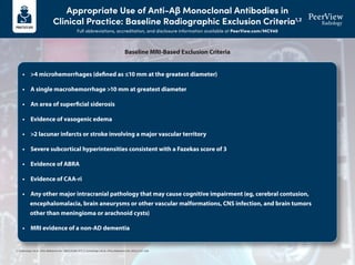
  • 1. Appropriate Use of Anti-Aβ Monoclonal Antibodies in Clinical Practice: Baseline Radiographic Exclusion Criteria1,2 Full abbreviations, accreditation, and disclosure information available at PeerView.com/MCV40 1. Cummings J et al. J Prev Alzheimers Dis. 2023;10:362-377. 2. Cummings J et al. J Prev Alzheimers Dis. 2022;2:221-230. • >4 microhemorrhages (defined as ≤10 mm at the greatest diameter) • A single macrohemorrhage >10 mm at greatest diameter • An area of superficial siderosis • Evidence of vasogenic edema • >2 lacunar infarcts or stroke involving a major vascular territory • Severe subcortical hyperintensities consistent with a Fazekas score of 3 • Evidence of ABRA • Evidence of CAA-ri • Any other major intracranial pathology that may cause cognitive impairment (eg, cerebral contusion, encephalomalacia, brain aneurysms or other vascular malformations, CNS infection, and brain tumors other than meningioma or arachnoid cysts) • MRI evidence of a non-AD dementia Baseline MRI-Based Exclusion Criteria
  • 2. Detecting, Monitoring, and Managing Amyloid-Related Imaging Abnormalities (ARIA) Full abbreviations, accreditation, and disclosure information available at PeerView.com/MCV40 3T scanner (recommended) 1.5T scanner (minimal) High field strength scanners have greater sensitivity but limited availability The use of 1.5T scanner is endorsed as a minimum standard Slice thickness: ≤5 mm Thinner slices increase resolution, but decrease signal-to-noise ratio TE: ≥20 ms Longer TE increases sensitivity to detection 2D T2* GRE or SWI (for ARIA-H) MRI sequences used to improve the detection, visualization, and monitoring of microhemorrhages and superficial siderosis (ARIA-H) T2-FLAIR (for ARIA-E) MRI sequences used to detect and monitor brain edema or sulcal effusion (ARIA-E) Diffusion-weighted imaging An MRI method used to differentiate between ARIA and cytotoxic processes (eg, infarcts) Recommended for differential diagnosis Visit the American Society for Neuroradiology (ASNR) website to access ARIA resources, including links to standardized protocols for 3T and 1.5T scanners via this QR code or URL: https://www.asnr.org/education-resources/alzheimers-webinar-series MRI Acquisition Protocols to Detect and Monitor ARIA1-3
  • 3. Detecting, Monitoring, and Managing Amyloid-Related Imaging Abnormalities (ARIA) Full abbreviations, accreditation, and disclosure information available at PeerView.com/MCV40 ARIA Type ARIA-E ARIA-H microhemorrhage ARIA-H superficial siderosis FLAIR hyperintensity confined to sulcus and/or cortex/subcortical white matter in one location <5 cm Mild Moderate Radiographic Severity Severe ≤4 new incident microhemorrhages One focal area of superficial siderosis FLAIR hyperintensity 5-10 cm, or more than one site of involvement, each measuring <10 cm 5-9 new incident microhemorrhages Two focal areas of superficial siderosis FLAIR hyperintensity measures >10 cm, often with significant subcortical white matter and/or sulcal involvement; ≥1 separate sites of involvement might be noted ≥10 new incident microhemorrhages >2 focal areas of superficial siderosis Classifying ARIA by Radiographic Severity4,5
  • 4. Detecting, Monitoring, and Managing Amyloid-Related Imaging Abnormalities (ARIA) Full abbreviations, accreditation, and disclosure information available at PeerView.com/MCV40 Headache Confusion and dizziness Neuropsychiatric symptoms Nausea Gait disturbance Visual disturbance/ blurred vision Seizure Less frequent Uncommon Symptom Severity Mild Discomfort noted; no disruption of daily activity Moderate Discomfort sufficient to reduce or affect normal daily activity Severe Incapacitating, with inability to perform normal daily activity Symptoms Consistent With ARIA That Should Trigger Out-of-Sequence MRI6-8
  • 5. Detecting, Monitoring, and Managing Amyloid-Related Imaging Abnormalities (ARIA) Full abbreviations, accreditation, and disclosure information available at PeerView.com/MCV40 Baseline MRI has no exclusion factors MRI routine or conducted because of symptoms suggestive of ARIA ARIA-E or ARIA-H detected Symptomatic Asymptomatic Radiographically mild ARIA-E or mild ARIA-H Continue treatment with anti-Aβ antibody; monthly MRI Continue treatment; discontinue monthly MRI if ARIA-E resolves or ARIA-H stabilizes Resume treatment with anti-Aβ antibody Suspend treatment; clinical assessment; repeat MRI monthly Radiographically moderate/severe ARIA-E or moderate/severe ARIA-H MRI shows resolution of ARIA-E or stabilization of ARIA-H; symptoms resolve; patient wishes to continue Stop anti-Aβ antibody therapy for any of the following • Any macrohemorrhage • >1 area of superficial siderosis • >10 microhemorrhages since treatment initiation • >2 episodes of ARIA • Severe symptoms of ARIA • Patient requires treatment with an anticoagulant Severity of Changes Observed on MRI Radiographic ARIA-E: Mild Radiographic ARIA-E: Moderate Radiographic ARIA-E: Severe Radiographic ARIA-H: Mild Radiographic ARIA-H: Moderate Radiographic ARIA-H: Severe Symptom Description No Symptoms Mild Symptoms Moderate Symptoms Severe Symptoms Discontinue dosing Discontinue dosing Discontinue dosing Discontinue dosing Discontinue dosing Discontinue dosing Discontinue dosing Discontinue dosing Discontinue dosing Discontinue dosing Suspend dosing Suspend dosing Suspend dosing Suspend dosing Suspend dosing Suspend dosing Suspend dosing Suspend dosing Suspend dosing Suspend dosing Discontinue dosing Discontinue dosing Continue dosing Continue dosing ARIA Management Algorithm6-8 1. Cogswell PM et al. AJNR Am J Neuroradiol. 2022;43:E19-E35. 2. Sperling RA et al. Alzheimers Dement. 2011;7:367-385. 3. Barakos J et al. J Prev Alzheimers Dis. 2022;9:211-220. 4. Aduhelm (aducanumab) Prescribing Information. https://www.accessdata.fda.gov/drugsatfda_docs/label/2023/761178s007lbl.pdf. 5. Leqembi (lecanemab) Prescribing Information. https://www.accessdata.fda.gov/drugsatfda_docs/label/2023/761269s000lbl.pdf. 6. Cummings J et al. J Prev Alzheimers Dis. 2023;10:362-377. 7. Cummings J et al. J Prev Alzheimers Dis. 2022;2:221-230. 8. Cummings J et al. J Prev Alzheimers Dis. 2021;4:398-410.
  • 6. Selected Images of Amyloid-Related Imaging Abnormalities (ARIA) and Its Mimics Full abbreviations, accreditation, and disclosure information available at PeerView.com/MCV40 Parenchymal Edema New T2-FLAIR hyperintense signal with mild local mass effect and sulcal effacement measuring <5 cm (mild ARIA-E) New multifocal, patchy T2-FLAIR hyperintense signal, each region measuring <5 cm (moderate ARIA-E); multiple ARIA-E yields a classification of moderate, as long as each region is <10 cm Extensive T2-FLAIR hyperintense signal throughout the right frontal and parietal lobes measuring >10 cm (severe ARIA-E); associated mass effect and sulcal effacement throughout much of the right hemisphere Baseline Baseline Baseline Post-treatment Post-treatment Post-treatment Mild Moderate Severe New sulcal T2-FLAIR hyperintense signal measuring <5 cm in transverse dimensions (mild ARIA-E) New T2-FLAIR sulcal effusion involving the right posterior temporal and parietal lobes measuring 5-10 cm (moderate ARIA-E) Extensive T2-FLAIR sulcal effusion involving the bilateral temporal and occipital lobes measuring ≥10 cm in extent (severe ARIA-E) Baseline Post-treatment Mild Sulcal Effusion Baseline Post-treatment Moderate Baseline Post-treatment Severe ARIA-E Examples (Detected With T2-FLAIR Sequence)1
  • 7. Selected Images of Amyloid-Related Imaging Abnormalities (ARIA) and Its Mimics Full abbreviations, accreditation, and disclosure information available at PeerView.com/MCV40 Microhemorrhage Postdosing, ≥10 new microhemorrhages (severe ARIA-H) Baseline Post-treatment Severe Postdosing, 5 treatment-emergent microhemorrhages (moderate ARIA-H) Baseline Post-treatment Moderate Postdosing, few (<5) new peripheral left frontal microhemorrhages (mild ARIA-H) Baseline Post-treatment Mild Superficial Siderosis Postdosing, new right temporal superficial siderosis, which involves contiguous sulci when viewed over multiple slices (mild ARIA-H, siderosis); this patient also had two treatment-emergent microhemorrhages (mild ARIA-H, microhemorrhage) Two regions of treatment-emergent superficial siderosis in the right greater-than-left frontal lobes (moderate ARIA-H) Baseline Post-treatment Mild Baseline Post-treatment Moderate ARIA-H Examples (Detected With T2-GRE or SWI Sequence)1
  • 8. Selected Images of Amyloid-Related Imaging Abnormalities (ARIA) and Its Mimics Full abbreviations, accreditation, and disclosure information available at PeerView.com/MCV40 Infarct (Early Subacute) FLAIR+ (hyperintensity) • In addition to FLAIR and GRE/SWI sequences, a trace DWI sequence should be included as routine protocol in ARIA monitoring examinations to help with the differential diagnosis of new signal abnormalities • For example, the DWI sequence plays an important role in helping to differentiate ARIA-E from potential cytotoxic edema caused by an incidental infarct • In classic cases of ARIA, the diffusion restriction will be absent, because intense diffusion restriction associated with an infarct is not a characteristic of ARIA • The DWI sequence helped to identify the underlying etiology of this patient’s radiographic findings • Diffusion restriction was identified, which indicated that it was not ARIA • However, the diffusion restriction was located in the sulci, which is not a typical pattern seen with acute or subacute infarcts • This patient was diagnosed with bacterial meningitis following confirmatory lab testing Infection (Bacterial Meningitis) FLAIR+ (vasogenic edema and sulcal effusions) GRE/SWI+ (microhemorrhages and superficial siderosis) DWI+ (with restricted diffusion in the sulci) DWI+ (with restricted diffusion) GRE/SWI+ (microhemorrhage) ARIA Mimics2
  • 9. Selected Images of Amyloid-Related Imaging Abnormalities (ARIA) and Its Mimics Full abbreviations, accreditation, and disclosure information available at PeerView.com/MCV40 1. Cogswell PM et al. Am J Neuroradiol. 2022;43:E19-E35. 2. Images courtesy of Tammie L.S. Benzinger, MD, PhD. ARIA Mimics2 Brain Metastasis FLAIR+ (hyperintensity) • New signal abnormalities detected in ARIA monitoring examinations may require T1-weighted gadolinium-enhanced imaging to differentiate between ARIA and brain metastases • Contrast-enhanced imaging should be considered based on the patient’s medical history and/or any clinical findings that may suggest the possibility of a metastatic etiology • ARIA and PRES are usually indistinguishable based on MRI (eg, both will typically have FLAIR hyperintensities and no restricted diffusion) • Therefore, the clinical context is critical to differentiate between ARIA and PRES • This patient presented to the emergency department with confusion, headache, and very high blood pressure (which is a classic presentation for PRES) • They were treated for their hypertension, which resolved their clinical symptoms and their edema Posterior Reversible Encephalopathy Syndrome (PRES) FLAIR+ (hyperintensities) GRE/SWI negative DWI negative (no diffusion restriction) T1 postcontrast + enhancement GRE/SWI+ (microhemorrhage) T1 postcontrast
  • 10. Use the following pages as reporting templates to document baseline screening MRI prior to initiating anti-amyloid therapy (page 1) and follow-up MRI to monitor ARIA in patients undergoing treatment with anti-amyloid therapy (pages 2-3). Printable Resource ARIA Reporting Templates Full abbreviations, accreditation, and disclosure information available at PeerView.com/MCV40
  • 11. Baseline MRI for Screening Prior to Initiating Anti-Amyloid Therapy (page 1 of 1) Scan Information (fill in or circle response as needed) DWI Gadolinium Contrast ARIA–H Sequence ARIA–E Sequence Slice Gap Slice Thickness (mm) Field Strength Model Manufacturer Y N Y N SWI GRE/T2* 3D T2-FLAIR 1.5T 3T Hyperintensities/Infarcts Summary Location (eg, frontal, parietal, occipital, temporal, brainstem, cerebellum) L/R Type (eg, parenchymal; sulcal) Max Diameter (cm) Lesion 1 Lesion 2 Lesion 3 Lesion 4 Microhemorrhages/Superficial Siderosis Summary Brainstem Deep Gray Cerebellum Temporal Frontal Parietal Occipital Total MCH count SS count Findings Total FLAIR hyperintensities: ______________ Describe locations and measure longest size in 1 dimension for each: _________________________________________________________________ Total microhemorrhages: ________________ Describe locations in general, deep vs lobar: ____________________________________________________________________________________ Total areas of superficial siderosis: __________________ Describe locations: _________________________________________________________________________________________________________ General description of other acute or chronic findings (eg, macrohemorrhage >10 mm; vasogenic edema; >2 lacunar infarcts or stroke involving a major vascular territory; severe subcortical hyperintensities consistent with a Fazekas score of 3; evidence of ABRA; evidence of CAA-ri; evidence of non-AD dementia; any other major intracranial pathology that may cause cognitive impairment): _____________________________________________________________________________________________________________________________ _____________________________________________________________________________________________________________________________ IMPRESSION Total microhemorrhages (circle one): 0-4 5-9 ≥10 Superficial siderosis (circle one): is not detected is present Note any other EXCLUSION CRITERIA for initiating treatment with anti-amyloid therapy: ____________________________________________________________________________________________________________________ Restricted Diffusion (circle correct response) Contrast Enhancement (circle correct response) Yes No N/A Yes No N/A Patient Information Patient name:____________________________ Age:________ Sex:_______ Patient ID:___________________ Referring MD:________________________ Report Information Scan Date:_________________________________ Site Location:_______________________________ Updated findings were conveyed to _____________________________ by _______________________________ (referring physician) (radiologist)
  • 12. Follow-Up MRI to Monitor for ARIA in Patients Undergoing Treatment With an Anti-Amyloid Therapy (page 1 of 2) Patient Information Patient name:____________________________ Age:________ Sex:_______ Patient ID:___________________ Referring MD:___________________________________________ Purpose of scan (circle correct response): scheduled/asymptomatic monitoring for ARIA OR unscheduled/safety MRI in response to symptoms Report Information Scan Date:________________________________ Site Location:______________________________ Scan Information (fill in or circle response as needed) DWI Gadolinium Contrast ARIA–H Sequence ARIA–E Sequence Slice Gap Slice Thickness (mm) Field Strength Model Manufacturer Y N Y N SWI GRE/T2* 3D T2-FLAIR 1.5T 3T ARIA-E Summary Location (eg, frontal, parietal, occipital, temporal, brainstem, cerebellum) L/R Type (eg, parenchymal; sulcal) Change From Baseline (cm) Lesion Dynamics (eg, new, enlarging, shrinking, stable Max Diameter (cm) Lesion 1 Lesion 2 Lesion 3 Lesion 4 Treatment History Anti-Amyloid Agent: ________________________________ Number of doses received: ____________ Date of last dose: _______________________ Suspected ARIA Symptoms (if present): ______________________ Restricted Diffusion (circle correct response) Contrast Enhancement (circle correct response) Yes No N/A Yes No N/A
  • 13. ARIA-H Summary Brainstem Deep Gray Cerebellum Temporal Frontal Parietal Occipital Total Current MCH count Baseline MCH count Change from baseline MCH count Current SS count Baseline SS count Change from baseline SS count Radiographic Severity (circle response) ARIA Type Severe Moderate Mild ≥1 location, >10 cm 1 location, 5-10 cm OR >1 location, each <10 cm 1 location, <5 cm ARIA-E ≥10 5-9 0-4 ARIA-H (MCH) >2 focal areas 2 focal areas 1 focal area ARIA-H (SS) Note other acute or chronic findings: _________________________________________________________________ _________________________________________________________________________________________________ Updated findings were conveyed to _____________________________ by _______________________________ (referring physician) (radiologist) Follow-Up MRI to Monitor for ARIA in Patients Undergoing Treatment With an Anti-Amyloid Therapy (page 2 of 2)
  • 14. ARIA Standardized Reporting Template for PowerScribe Full abbreviations, accreditation, and disclosure information available at PeerView.com/MCV40 Download the XML code for a standardized and automated ARIA reporting template in PowerScribe by scanning the QR code or visiting the provided URL: www.PeerView.com/ARIA24-PowerScribeXML Once the file is downloaded, the code will need to be updated for your radiology department.