Stages of Amelogenesis and Enamel Formation
•Transferir como PPTX, PDF•
1 gostou•2,697 visualizações

Development of enamel is an unique process

Recomendados
Mais conteúdo relacionado
Mais procurados
Mais procurados (20)
Semelhante a Stages of Amelogenesis and Enamel Formation
Semelhante a Stages of Amelogenesis and Enamel Formation (20)
Mais de sherifsayed65
Mais de sherifsayed65 (9)
Último
Último (20)
Stages of Amelogenesis and Enamel Formation
- 1. Amelogenesis
- 2. Objectives of studying amelogenesis 1. Understand the nature of enamel. 2. Appreciate how this dental tissue is unique. 3. Appreciate how the enamel protect the underlying dentin. 4. Appreciate the physical properties of enamel. 5. Know the chemical composition of enamel.
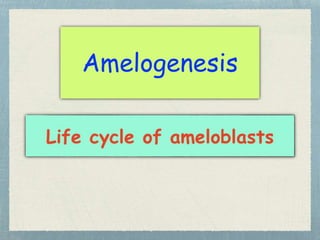
- 3. 1. Morphogenic stage. 2. Differentiating stage (Organizing stage). 3. Secretory stage (Formative stage). 4. Maturative stage (Calcifying stage). 5. Protective stage. 6. Desmolytic stage. Life cycle of ameloblasts
- 4. 1-Morphogenic stage of IEE at cap stage Structure of inner enamel epithelium : 1.Columnar cells separated from dental papilla by basement membrane. 2.Large centrally located nucleus. 3.Proximal end: The end of IEE facing stellate reticulum. 4.Distal end: The end of the Inner dental epithelium facing dental papilla. 5.Active cells contain RER,GC, ribosomes and mitochondria. IEE proximal end Distal end BM
- 5. 2-Differentiating stage of IEE at early bell stage • Differentiation: 1. The cell become tall columnar leading to disappearance of cell free zone. 2. Nucleus move toward proximal end. 3. Mitochondria move toward proximal end. 4. GC & RER move toward the distal end. 5. Ameloblast begin to secrete the organic matrix of enamel. 6. Ameloblast cells are attached together by desmosome termed web which are two types: (A) Proximal terminal web at the proximal end. (B) Distal terminal web at the distal end. DTW PTW
- 6. 3-Secretory stage of ameloblasts Enamel matrix Odontoblast cells Dentin matrix Ameloblast cells PTW Enamel matrix formation and secretion 1. Ribosomes out from the nucleus to RER in the cytoplasm. 2. protein is formed inside RER according to the amino code of ribosomes. 3. RER form small granule contain protein. 4. This small granule go to GC to add CHO and form secretory granules contain enamel matrix and out to the cytoplasm. 5. This granule go toward the distal end of the ameloblast and get out from the cell in the front of newly formed dentin.
- 7. • Tome’s process 1. After formation of the first layer of enamel matrix, the ameloblast move away from the dentin to allow for further enamel matrix deposition. 2. By this movement , each ameloblast develop conical process termed tomes process. 3. Tom's process are demarcated from cell body by distal terminal web. 4. Tom's process contain secretory granules. 5. The rest of enamel are secreted from tom's process. Tome’s process
- 8. .
- 9. Pulp Dentin and enamel firstly formed at cusp tip or incisal edge and then sweep down the slopes toward cervical line.
- 10. 4- Maturative stage (calcification stage) striated border 1.Ameloblast reduced in length. 2.Decrease of number of cell organelles. 3.Disappearance of tom's processes. 4.Distal end of the cells become ruffled and termed ruffled border. 5.The become ruffled engulf the enamel matrix with secretion of calcium and phosphorus for enamel calcification, so, enamel become mature.
- 11. 5- Protective stage Primary enamel cuticle After complete maturation of enamel , ameloblast lose their ruffled border and secrete thin organic layer on the crown termed primary enamel cuticle. Reduced Enamel Epithelium • Ameloblast become flat and lose their regular arrangement and can not be differentiated from the other cells of dental organ. • All cells of dental organ form a layer cover the crown termed reduced dental epithelium which protect the crown from the surrounding connective tissue. • If the reduced dental epithelium destroyed from above the crown, the enamel may be resorbed by the action of connective tissue cells.
- 12. • During process of tooth eruption to the oral cavity, the reduced dental epithelium proliferate by cell division and secrete enzyme for destruction of connective tissue separating it from oral epithelium. • This enzyme is termed desmolytic enzyme. Then, the reduced enamel epithelium fused with the oral epithelium and become part of gingiva during tooth erupt. 6- Desmolytic stage
- 13. Amelogenesis Stage of enamel formation 1. Stage of organic matrix formation of enamel. 2. Stage of mineralization and maturation of enamel. (A) Immediate partial mineralization up to 25-30%. (B) Maturation up to 96%.
- 14. Finish