Chest X-Ray Diagnosis of Cardiac Conditions
•Transferir como PPTX, PDF•
26 gostaram•3,570 visualizações

The document describes various congenital heart defects categorized into 5 groups based on their appearance on chest x-ray. Group I lesions show increased pulmonary blood flow without cyanosis. Group II lesions cause cyanosis with decreased lung vascularity and normal heart size. Group III lesions also cause cyanosis but with decreased lung vascularity and enlarged heart. Group IV lesions have increased pulmonary blood flow causing cyanosis. Group V shows signs of pulmonary venous congestion. Specific defects are described within each group along with their characteristic chest x-ray findings.

Recomendados
Recomendados
Mais conteúdo relacionado
Mais procurados
Mais procurados (20)
Semelhante a Chest X-Ray Diagnosis of Cardiac Conditions
Semelhante a Chest X-Ray Diagnosis of Cardiac Conditions (20)
Mais de Malleswara rao Dangeti
Mais de Malleswara rao Dangeti (20)
Último
Último (20)
Chest X-Ray Diagnosis of Cardiac Conditions
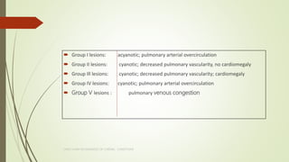
- 1. Group I lesions: acyanotic; pulmonary arterial overcirculation Group II lesions: cyanotic; decreased pulmonary vascularity, no cardiomegaly Group Ill lesions: cyanotic; decreased pulmonary vascularity; cardiomegaly Group IV lesions: cyanotic; pulmonary arterial overcirculation Group V lesions : pulmonary venous congestion CHEST X RAY IN DIAGNOSIS OF CARDIAC CONDITIONS
- 2. Group I lesions: acyanotic; pulmonary arterial overcirculation ASD PAPVC AVCD(endocardial cushion defect) VSD PDA Other aortic level shunts (e.g., RSOV, AP window) CHEST X RAY IN DIAGNOSIS OF CARDIAC CONDITIONS
- 3. Group II lesions: cyanotic; decreased pulmonary vascularity, no cardiomegaly TOF Transposition with pulmonic stenosis and VSD DORV with PS and VSD DOLV with PS and VSD Single ventricle with PS CCTGA with PS and VSD Pulmonic atresia with intact ventricular septum, type I Some types of tricuspid atresia (large ASD and pulmonary stenosis or atresia)CHEST X RAY IN DIAGNOSIS OF CARDIAC CONDITIONS
- 4. Group Ill lesions: cyanotic; decreased pulmonary vascularity; cardiomegaly Ebstein's anomaly PA (critical) with ASD or PFO Some types of tricuspid atresia (restrictive ASD) Pulmonary atresia with intact ventricular septum, type II transient TR of the newborn CHEST X RAY IN DIAGNOSIS OF CARDIAC CONDITIONS
- 5. Group IV lesions: cyanotic; pulmonary arterial overcirculation TGA without PS Truncus arteriosus TAPVC-non obstrcutive Tricuspid atresia without PS Single ventricle without PS DORV without PS Double-outlet left ventricle Pulmonary AV fistulae CHEST X RAY IN DIAGNOSIS OF CARDIAC CONDITIONS
- 6. Group V -pulmonary venous congestion Critical CoA Critical AS TAPVC-obstructive CHEST X RAY IN DIAGNOSIS OF CARDIAC CONDITIONS
- 7. CHEST X RAY IN DIAGNOSIS OF CARDIAC CONDITIONS
- 8. ATRIAL SEPTAL DEFECT RAE,RVE Dilation of RVOT -smooth continuity with enlarged PT above OS ASD RVE, RAE . LV is hypovolaemic and hypoplastic. RPA is more prominent than LPA giving radiological sign of jug- handle appearance. OP ASD LV enlargement also SVC type SINUS VENOSUS ASD SUBTLE LOCALIZED DILATION OF SVC AS IT JOINS RA CHEST X RAY IN DIAGNOSIS OF CARDIAC CONDITIONS
- 9. ASD CHEST X RAY IN DIAGNOSIS OF CARDIAC CONDITIONS
- 10. 1. Pulmonary vascularity –decreased but peripheral pruning does not occur PT and its right branch are dilated and contain eggshell calcium (Ca). RA is enlarged, and a dilated RV occupies apex. Cardiomegaly persist despite devolepment of ES CHEST X RAY IN DIAGNOSIS OF CARDIAC CONDITIONS
- 11. VSD Cardiomegaly : proportional to volume overload LV, LA and RV enlargement ascending aorta is inconspicuous. Both PAs are equally prominent. Rt aortic arch-2% Large shunts in infants - hyperinflated lungs with flat hemidiaphragms CHEST X RAY IN DIAGNOSIS OF CARDIAC CONDITIONS
- 12. appreciable enlargement of LV in context of no more than a modest left-to-right shunt ascending aorta is prominent and pulsates vigorously on fluoroscopy. perimembranous or subarterial VSD- onset of AR is insidious, so x-ray initially reflects left-to-right shunt. As time goes on, balance shifts CHEST X RAY IN DIAGNOSIS OF CARDIAC CONDITIONS
- 13. AVCD 11 ribs, double manubrial ossification ,tall vertebral bodies When left AV valve regurgitation coexists, RA is especially enlarged because regurgitant flow is directed into RA cavity left cardiac border is straightened by a prominent RVOT Dilated RA occupies right lower cardiac border, and LV can occupy apex despite RV enlargement Dilated PT may be eclipsed by a prominent RVOT ascending aorta is inconspicuous CHEST X RAY IN DIAGNOSIS OF CARDIAC CONDITIONS
- 14. CHEST X RAY IN DIAGNOSIS OF CARDIAC CONDITIONS
- 15. lateral x-ray in Down syndrome - double manubrial ossification center PA projection consistently discloses an absent or rudimentary 12th rib Hyperinflation of lungs = upper airway obstruction in Down syndrome flattens hemodiaphragms CHEST X RAY IN DIAGNOSIS OF CARDIAC CONDITIONS
- 16. disproportionate RA enlargement cardiac silhouette occasionally has a ball-like shape right side of ball -large RA left side of ball -dilation of RV infundibulum and LV CHEST X RAY IN DIAGNOSIS OF CARDIAC CONDITIONS
- 17. LA LV RA AO PT VSD ↑ ↑ ↔ ↓ ↑ AVCD ↑ ↑ ↑ ↓ ↑ VSD - AR ↔ ↑ ↑ ↔ ↑ ↔ ↑ GERBODE ↑ ↑ ↑ ↓ ↑ CHEST X RAY IN DIAGNOSIS OF CARDIAC CONDITIONS
- 18. PATENT DUCTUS ARTERIOSUS Enlargement of left heart chambers. Enlargement of ascending aorta or aortic arch. Pulmonary plethora with enlarged central and peripheral artery. Filling up of angle between aortic arch and PA(radiological AP Window): Most specific sign. • Possible PDA calcification in adults(inverted Y shaped ) Both PAs are equally dilated CHEST X RAY IN DIAGNOSIS OF CARDIAC CONDITIONS
- 19. CHEST X RAY IN DIAGNOSIS OF CARDIAC CONDITIONS
- 20. PAPVC Pulmonary arterial overcirculation: This may be apparent or more severe only in the lung with anomalous drainage. RAE RVE Enlargement of main and hilar pulmonary arterial segments Small ascending aorta and aortic arch Enlargement of SVC, azygous vein, CS or other systemic veins, depending on site of connection Prominent LSVC Abnormal course of pulmonary veins through the lung or in relation to mediastinal margins CHEST X RAY IN DIAGNOSIS OF CARDIAC CONDITIONS
- 21. SCIMITAR Syndrome CHEST X RAY IN DIAGNOSIS OF CARDIAC CONDITIONS • scimitar sign • RPA is hypolplasic-small right hilar shadow Dextroposition of heart Hypoplasia of right lung
- 22. CHEST X RAY IN DIAGNOSIS OF CARDIAC CONDITIONS
- 23. TETROLOGY OF FALLOT Decreased pulmonary vascularity -Normal vascularity in a cyanotic individual is equated with decreased vasculariy since the distinction between normal and mildly decreased vascularity is frequently difficult. Normal or nearly normal cardiac size shape of a wooden shoe or boot (in French, coeur en sabot) = uplifting of cardiac apex due to RVH and concavity of MPA Concave or absent MPA Small hilar pulmonary arteries: This may be most evident on the lateral view. Dilated ascending aorta right aortic arch = 25% . Asymmetric pulmonary vascularity is frequent, especially because of associated branch PA stenosis. Lsvc shadow Unilateral rib notching after BT shunt CHEST X RAY IN DIAGNOSIS OF CARDIAC CONDITIONS
- 24. CHEST X RAY IN DIAGNOSIS OF CARDIAC CONDITIONS
- 25. TOF with pulmonary atresia Bilateral reticular formation -bronchopulmonary collaterals-Lacy reticular pattern without the normal diminution in vessel caliber toward the periphery because systemic arterial collaterals anastomose with segmental or lobar intrapulmonary arteries When systemic arterial collaterals or bronchial collaterals anastomose with hilar or extrapulmonary arteries, intrapulmonary branching is normal patterns of collateral arterial circulation are not uniform, with some areas oligemic and others normal or hypervascular Systemic collateral arteries rarely cause rib notching because they do not run in intercostal grooves. Cardiac size tends to be larger in response to flow through systemic arterial collaterals reticular lacy appearance
- 26. CHEST X RAY IN DIAGNOSIS OF CARDIAC CONDITIONS
- 27. TOF with absent pulmonary valve Decreased distal pulmonary vascularity Cardiac size variable, depending on severity of PR Infundibular dilation projects leftward as a hump- shaped shadow dilated RV occupies the apex enlarged RA Pulmonary vascularity is normal rather than decreased. CHEST X RAY IN DIAGNOSIS OF CARDIAC CONDITIONS Aneurysmal enlargement of main and central pulmonary arteries
- 28. CHEST X RAY IN DIAGNOSIS OF CARDIAC CONDITIONS
- 29. EBSTEINS ANOMALY cardiac silhouette • near normal to diagnostic • Heart size in symptomatic infants is immense Pulmonary vascularity • normal in mild acyanotic Ebstein’s • reduced when anomaly is severe and cyanotic . infundibulum • straightens left cardiac border or forms a conspicuous convex shoulder RA silhouette • almost always enlarged . • seldom normal even when cardiac silhouette is normal CHEST X RAY IN DIAGNOSIS OF CARDIAC CONDITIONS
- 30. EBSTEINS ANOMALY BOX SHAPED HEART • Marked rightward convexity of enlarged RA + marked leftward convexity of enlarged infundibulum . vascular pedicle • narrow because PT is not border forming and ascending aortic shadow is inconspicuous or absent . • resembles pericardial effusion CHEST X RAY IN DIAGNOSIS OF CARDIAC CONDITIONS
- 31. EBSTEINS ANOMALY CHEST X RAY IN DIAGNOSIS OF CARDIAC CONDITIONS Result of marked angulation at superior vena caval- RA junction as RA enlarges.
- 32. PA WITH INTACT IVS TYPE 1 cardiac silhouette at birth may be normal . PA segment is normal because PT develops normally despite PV atresia ascending aorta is enlarged RA shadow is moderately prominent well formed convex LV occupies apex TYPE 2 cardiac silhouette virtually fills chest—“wall-to wall”—remarkable enlargement of RA and RV CHEST X RAY IN DIAGNOSIS OF CARDIAC CONDITIONS
- 33. CHEST X RAY IN DIAGNOSIS OF CARDIAC CONDITIONS MPA is normal ascending aorta (Ao) is enlarged
- 34. CHEST X RAY IN DIAGNOSIS OF CARDIAC CONDITIONS
- 35. TA restrictive VSD and NRGA(type 1b) • right cardiac contour – distinctive ,enlargement of RA and its appendage and accentuated by a flat receding inferior border that reflects absence of RV • Hump like on right cardiac border • convex LV occupies apex • Pulmonary vascularity is reduced • MPA is inconspicuous • ascending aorta -prominent • Normal vascular pedicle • Rt aortic arch-5-8% intact ventricular septum(type 1a) • ascending aorta is more conspicuous • lung fields -lacy vascular pattern of systemic arterial collateralsCHEST X RAY IN DIAGNOSIS OF CARDIAC CONDITIONS
- 36. CHEST X RAY IN DIAGNOSIS OF CARDIAC CONDITIONS large LV occupies the apex Pulmonary vascularity is reduced ascending aorta (Ao) is prominent MPA is inconspicuous dilated RA recedes acutely because of absence of RV
- 37. VSD is nonrestrictive and PVR is low(1c) • pulmonary vascularity is increased • PT and LA and RA are enlarged • prominent LV is apex-forming tricuspid atresia with complete TGA, a nonrestrictive VSD , and low PVR(II c) • increased pulmonary vascularity • Inconspicuous Aorta • enlargement of LA and RA( CARDIOMEGALY) • prominent apex-forming LV. • vascular pedicle is narrow • resembles uncomplicated complete TGA • Left cardiac border is straight if there is left-sided juxtaposition of atrial appendages. PVR is increased • lungs are oligemic • heart size is normal or nearly soCHEST X RAY IN DIAGNOSIS OF CARDIAC CONDITIONS
- 38. TA + TGA + PS(type 2b) normal or reduced pulmonary vascularity prominent RA Convex LV narrow vascular pedicle juxtaposition of the atrial appendages pointed bulge on the left border of the mediastinal shadow below the region where PA should be seen right border of RA is straight because the absence of RV pulls the border toward RA.
- 39. CHEST X RAY IN DIAGNOSIS OF CARDIAC CONDITIONS JUXTRA POSITION
- 40. Pulmonary oligaemia Normal vascular pedicle 1B Narrow vascular pedicle II B Pulmonary plethora Normal vascular pedicle IC Narrow vascular pedicle IIC Lacy reticular pattern 1A CHEST X RAY IN DIAGNOSIS OF CARDIAC CONDITIONS
- 41. CHEST X RAY IN DIAGNOSIS OF CARDIAC CONDITIONS
- 42. TRANSPOSITION OF GREAT ARTERIES RA border is abnormally convex LA is enlarged because of increased PBF. egg on its side. Increased CT ratio with egg lying on its side appearance blunter right border of egg =RA convex left border –LV CHEST X RAY IN DIAGNOSIS OF CARDIAC CONDITIONS
- 43. D-TGA ,VSD without PS neonatal x-ray -Normal typical features –once PVR falls and PBF increases Increased pulmonary vascularity distribution of PBF favors right lung because of rightward direction of PT A progressive increase in flow into right lung may culminate in a substantial decrease in flow to left crural portions of hemidiaphragms are low when lungs are hyperinflated CHEST X RAY IN DIAGNOSIS OF CARDIAC CONDITIONS D T G A
- 44. Thymic shadow is almost always absent after first 12 hours of life narrow vascular pedicle -PT is posterior and medial pedicle is narrowest when ascending aorta courses vertically upward directly anterior to PT aortic root is seldom sufficiently rightward to be border forming except when ascending aorta enlarges in presence of leftward and posterior malalignment of infundibular septum CHEST X RAY IN DIAGNOSIS OF CARDIAC CONDITIONS D T G A
- 45. vascular pedicle - widens when a dilated hypertensive posterior PT is convex to left When PS and VSD coexist- right aortic arch is present in 11% to 16% lateral projection= heart assumes a circular appearance because an enlarged RV merges with anterior aorta, and an enlarged LV merges with dilated posterior LA; widened vascular pedicle Discrepency between pulm plethora & hilar and main pulm arteries due to midline position of PT CHEST X RAY IN DIAGNOSIS OF CARDIAC CONDITIONS D T G A
- 46. Juxtaposition of atrial appendages localized bulge along mid left cardiac border that represents contiguous mass of two appendages ↑ PVR lung vascularity decreases, size of LV and LA decrease, and a dilated hypertensive posterior PT emerges at left base Severe fixed PS decreased pulmonary vascularity, a small LV, a small LA, and enlargement of RV and RA CHEST X RAY IN DIAGNOSIS OF CARDIAC CONDITIONS D T G A
- 47. D TGA CHEST X RAY IN DIAGNOSIS OF CARDIAC CONDITIONS Narrow vasc pedicle RA border is abnormally convex Increased pulmonary vascularity egg on its side. Thymic shadow is absent
- 48. All four chambers are dilated with pulmonary plethora. Left side chamber enlargement > right side chamber enlargement Enlargement of RV and RA –CCF /incompetent biventricular truncal valve. 1/3 rd of cases right aortic arch . large truncus arteriosus resembles a large ascending aorta that may continue as a right aortic arch (35%) and a high transverse aortaCHEST X RAY IN DIAGNOSIS OF CARDIAC CONDITIONS
- 49. ↑ pulmonary vascularity MPA segment is prominent (type 1) MPA segment is absent (type 2 or 3)-concave profile -RAO prominent LPA may reveal itself as a high shadow that curves upward to form a left hilar ( especially evident in right aortic arch)-HILAR COMMA SIGN convex MPA segment of truncus type 1 tends to arise at a higher level compared with other forms of PA dilation arterial pedicle appear narrow discrepancy between the vascular markings on the two sides -unilateral atresia, or absence, of one PA Absence of PA is usually on same side as aortic arch in contrast to TOF CHEST X RAY IN DIAGNOSIS OF CARDIAC CONDITIONS t r u n c u s a r t e r i o s u s
- 50. older adults with PAH Dilated hypertensive MPA segment is especially prominent ↓ pulmonary vascularity increased prominence of MPA and its right and left branches relatively normal LV CHEST X RAY IN DIAGNOSIS OF CARDIAC CONDITIONS t r u n c u s a r t e r i o s u s
- 51. CHEST X RAY IN DIAGNOSIS OF CARDIAC CONDITIONS snowman or figure of eight appearance. Supracardiac type to LIV,mixed variety(<1/3rd cases) head of snowman dilated vertical vein on left innominate vein on top SVC on right body enlarged RA
- 52. Without PVO ↑ PBF RA,RV,PA- enlarged With PVO Pulm venous HTN signs No cardiomegaly Reticular pattern Kerly b lines Ground glass opacities CHEST X RAY IN DIAGNOSIS OF CARDIAC CONDITIONS
- 53. snowman or figure of eight appearance Develops once PVR falls(3-6 months) VSD associated with large THYMUS- Psudo snowman appearance Anamalous drainage to RSVC-bulge on right upper mediastinus Aortic knob-small. CHEST X RAY IN DIAGNOSIS OF CARDIAC CONDITIONS
- 54. TAPVR CHEST X RAY IN DIAGNOSIS OF CARDIAC CONDITIONS
- 55. CHEST X RAY IN DIAGNOSIS OF CARDIAC CONDITIONS
- 56. DOUBLE OUTLET RV Generalized cardiomegaly increased pulmonary vascularity. aorta and PA have a more side-to-side configuration-cardiac waist is relatively wider CHEST X RAY IN DIAGNOSIS OF CARDIAC CONDITIONS
- 57. DORV WITH SUB AORTIC VSD(no PS) CHEST X RAY IN DIAGNOSIS OF CARDIAC CONDITIONS low PVR ~ non-restrictive perimembranous VSD with ↑ PBF Thymus is present even though there is transposition of aorta PT is prominent because it carries increased volume at systemic pressure and is not posterior to aorta LA and LV enlargement CCF= RA and RV dilate elevated PVR (before neonatal fall or after development of PVD) lung fields are oligemic RV copes with systemic resistance without enlarging significantly ~non-restrictive VSD and ES
- 58. CHEST X RAY IN DIAGNOSIS OF CARDIAC CONDITIONS pulmonary vascularity is increased prominent RA dilated LV occupies the apex PT is moderately convex
- 59. DORV WITH SUBAORTIC VSD AND PS (resembles TOF) PT is not dilated When PS is mild, pulmonary vascularity is increased and LV is dilated When PS is severe, pulmonary vascularity is reduced, heart size is normal, and apex is convex pulmonary atresia= ascending aorta is enlarged, MPA is concave, and apex is boot-shaped. CHEST X RAY IN DIAGNOSIS OF CARDIAC CONDITIONS
- 60. DORV WITH SUBPULMONARY VSD: TAUSSIG-BING ANOMALY increase in pulmonary arterial and pulmonary venous vascularity results from low PVR and CCF LA and LV are enlarged RA and RV are enlarged - CCF dilated PT projects prominently to left when great arteries are side-by- side-resembles a nonrestrictive perimembranous VSD When dilated PT is posterior and therefore not border-forming, x-ray resembles D TGA except for presence of a thymus CHEST X RAY IN DIAGNOSIS OF CARDIAC CONDITIONS
- 61. CHEST X RAY IN DIAGNOSIS OF CARDIAC CONDITIONS Pulmonary vascularity is increased thymus enlarged RA
- 62. ↑ PVR PBF decreases pulmonary venous vascularity disappears Volume overload of LV is curtailed, but dilation of PT persists resembles a nonrestrictive VSD with ES CHEST X RAY IN DIAGNOSIS OF CARDIAC CONDITIONS
- 63. SV inverted outlet chamber localized convexity at upper left cardiac border aorta -convex to left or rises vertically as in CCTGA A transposed posteromedial PT may lift its dilated right branch and create a waterfall appearance Holmes heart inverted outlet chamber is distinctively convex concordant PT noninverted outlet chamber aorta -convex to right but is not border-forming as is case in D-TGA Narrow vasc pedicle Thymus present CHEST X RAY IN DIAGNOSIS OF CARDIAC CONDITION S
- 64. single morphologic left ventriclewith inverted outlet chamber CHEST X RAY IN DIAGNOSIS OF CARDIAC CONDITIONS OC forms a convex bulge and gives rise to the aorta Ao SV and RA are dilated
- 65. single morphologic left ventricle and a noninverted outlet chamber CHEST X RAY IN DIAGNOSIS OF CARDIAC CONDITIONS Dilated right atrium (RA) narrow vascular pedicle
- 66. SV With exception of Holmes heart great arteries are transposed ( aorta from OC and PT from m LV) size of cardiac silhouette increases -excessive PBF and volume overload of SV LAE-lateral films or with a barium esophagram because what appears to be LA appendage in PA projection is an inverted OC RA dilation- CCF, which is reinforced by subaortic stenosis CHEST X RAY IN DIAGNOSIS OF CARDIAC CONDITIONS
- 67. SV single Mlv with severe PS inverted outlet chamber size of heart is normal inverted outlet chamber -bulge at left upper cardiac border dilated aorta that arises from an inverted outlet chamber - convexity to left or that ascends vertically and is not border- forming on either side Pulmonary atresia with an inverted outlet chamber box-like cardiac silhouette dilated ascending aorta forming left upper border that merges with a small underfilled ventricle below and vertebral column forming straight right border CHEST X RAY IN DIAGNOSIS OF CARDIAC CONDITIONS
- 68. single morphologic lv and severe PS noninverted outlet chamber CHEST X RAY IN DIAGNOSIS OF CARDIAC CONDITIONS No cardiomegaly.
- 69. Holms Heart CHEST X RAY IN DIAGNOSIS OF CARDIAC CONDITIONS
- 70. SV-univentricular hearts of RV morphology both GAs necessarily arise from single RV a form of DORV vascular pedicle is narrow -aorta is anterior and PT is posterior or wide if side-by-side. PS is common pulmonary vascularity is normal or reduced heart size is not significantly increased. CHEST X RAY IN DIAGNOSIS OF CARDIAC CONDITIONS
- 71. CCTGA normal - distinctive triad of contours that consists of ascending aorta on right and aortic knuckle and PT on left CCTGA- this triad is lost because aorta does not ascend on right and PT is not border forming on left MC relationship ascending aorta -anterior and to left medially and posteriorly positioned PT ascending aorta at left base varies from absent to straight to gently concave to moderately convex to strikingly convex CHEST X RAY IN DIAGNOSIS OF CARDIAC CONDITIONS C C T G A
- 72. CCTGA Less commonly , ascending aorta rises vertically and anterior to posterior PT, so neither great artery is border forming posterior and rightward PT tilts right branch upward and left branch downward, so that two branches are at same level dilated posterior PT can displace SVC to right, forming a right basal shadow or may project as a right basal convexity that can be mistaken for ascending aorta CHEST X RAY IN DIAGNOSIS OF CARDIAC CONDITIONS C C T G A
- 73. CCTGA silhouette of mRV : (1) a hump-shaped appearance (prominent inverted infundibulum ) (2) a septal notch(subtle indentation just above left hemidiaphragm corresponding to apical position of interventricular groove ) hump-shaped infundibular shadow occupies site of left atrial appendage so LAE is best identified in a lateral projection. giant LA- huge ball suspended below a narrow vascular pedicle ( due to left AV valve regurgitation or Ebstein anamoly or ↑ PBF ) CHEST X RAY IN DIAGNOSIS OF CARDIAC CONDITIONS C C T G A
- 74. L TGA RPA high take-off because of an absent aortic shadow and is also quite prominent waterfall" appearance - prominence of Rt pulmonary hilum with displacement medially and superiorly. left heart border boxlike, straightened Dextrocardia usually occurs with normal abdominal situs (20 percent of ccTGA ) abdominal situs solitus and dextrocardia should raise suspicion of ccTGA. CHEST X RAY IN DIAGNOSIS OF CARDIAC CONDITIONS C C T G A
- 75. CCTGA , a nonrestrictive VSD, and increased PBF septal notch dilated posterior PT causes rightward displacement of SVC
- 76. Normal pulmonary vascularity Normal cardiac size RVE: This is usually detected initially on the lateral view as a prominent convexity of the anterior cardiac border or filling of the retrosternal space. Poststenotic dilatation of MPA Dilated and usually laterally displaced LPA CHEST X RAY IN DIAGNOSIS OF CARDIAC CONDITIONS
- 77. CHEST X RAY IN DIAGNOSIS OF CARDIAC CONDITIONS OLIGEMIA CARDIOMEGALY RAE,RVE DILATED MPA CRITICAL PS
- 78. CONG AS neonates with severe AS = pulmonary venous congestion /Pulmonary edema Cardiomegaly LVE CHEST X RAY IN DIAGNOSIS OF CARDIAC CONDITIONS
- 79. AS LEVEL of stenosis • Calcification (stenosis is valvular) • Size of aorta SEVERITY • dense calcium • increase in LA size LV • remain normal-sized through a wide range of severity • adaptive response is concentric hypertrophy with a normal or reduced cavity • enlarges downward and to left and posterior • frontal view -apex extends below left hemidiaphragm • Left lateral projection-extends behind IVC CHEST X RAY IN DIAGNOSIS OF CARDIAC CONDITIONS
- 80. CHEST X RAY IN DIAGNOSIS OF CARDIAC CONDITIONS Valvular AS Subvalvular AS
- 81. Significant LV enlargement infants - severe AS and CCF adults = CCF, whether or not AS is severe CHEST X RAY IN DIAGNOSIS OF CARDIAC CONDITIONS
- 82. ascending aorta Dilation bicuspid AS Turner’s syndrome normal fixed subvalvular aortic stenosis 50%-dilated Aorta normal to small supravalvular AS undersized hypoplasia of ascending aorta hypoplastic aortic annulus and a miniature valve CHEST X RAY IN DIAGNOSIS OF CARDIAC CONDITIONS
- 83. CoA infants asymptomatic x-ray is normal symptomatic infants pulmonary venous congestion with dilation of RV and RA and LA LV size remains normal children and young adults postcoarctation descending thoracic aorta has a distinctive leftward convexity + dilation of LSCA Rib notching CHEST X RAY IN DIAGNOSIS OF CARDIAC CONDITIONS
- 84. CRITICAL COA pulmonary venous hypertension / pulmonary edema Cardiomegaly LV enlargement No rib notching/aortic knob is not characterstic Pulmonary plethora-VSD/PDA CHEST X RAY IN DIAGNOSIS OF CARDIAC CONDITIONS
- 85. Notching of ribs collateral flow through dilated, tortuous, pulsatile posterior intercostal arteries Notches vary from rib to rib and from patient to patient and may be single, multiple, shallow, deep, broad, or narrow. anterior ribs are spared because anterior intercostal arteries do not run in intercostal grooves. Rib notching seldom appears before age 6 years Lateral x-rays = retrosternal notching or scalloping caused by dilated tortuous IMA CHEST X RAY IN DIAGNOSIS OF CARDIAC CONDITIONS
- 86. • bilateral notching between third & eighth ribsCoA distal to LSCA • unilateral rib notching of right hemithorax LSCA lumen is compromised • unilateral notching of left hemithorax. Anomalous origin of RSCA distal to coarctation • confined to lower ribsabdominal CoA CHEST X RAY IN DIAGNOSIS OF CARDIAC CONDITIONS
- 87. older children and adults = ascending aorta is moderately to markedly dilated Turner’s syndrome-ascending aorta may be aneurysmal Proximal and distal paracoarctation aorta -dilated calcification is occasionally visible in wall of aneurysm. dilated LSCA proximal to coarctation and a dilated aorta distal to coarctation -figure 3 silhouette mirror image of figure three sign - barium esophagram LSCA cannot dilate when its lumen is compromised by coarctation, so dilation of distal paracoarctation aorta exists alone CHEST X RAY IN DIAGNOSIS OF CARDIAC CONDITIONS
- 88. CHEST X RAY IN DIAGNOSIS OF CARDIAC CONDITIONS figure 3 silhouette CoA that obstructed the orifice of LSCA which is therefore not dilated unilateral notching of the ribs ascending aorta (AAo) is dilated
- 89. Coarctation of aorta LAO , obtained with barium esophagography CHEST X RAY IN DIAGNOSIS OF CARDIAC CONDITIONS
- 90. irregular scalloped notching of inferior margins of posterior ribs CHEST X RAY IN DIAGNOSIS OF CARDIAC CONDITIONS
- 91. CHEST X RAY IN DIAGNOSIS OF CARDIAC CONDITIONS Aortic obstruction • COA • IAA • Acquired obstruction of the aorta: Takayasu's aortitis, atherosclerotic obstruction, • Unusual causes of coarctation: neurofibromatosis, Williams' syndrome, rubella syndrome Subclavian arterial obstruction • BT shunt (upper 2 ribs) • Takayasu's arteritis (usually unilateral) • Atherosclerosis Severely reduced pulmonary blood flow • TOF • Pulmonary atresia • Tricuspid atresia • Unilateral absence or atresia of a PA • Pulmonary emphysema • Chronic pulmonary thromboernbolic disease • Superior vena canal obstruction Vascular shunts • Pulmonary AV shunt • Intercostal AV shunt • Intercostal to PA shunt • Intercostal neuroma Poliomyelitis (upper margin) • Hyperparathyroiclism
- 92. retroesophageal aberrant RSCA -posterior indentation of barium esophagram kinked aorta of pseudocoarctation transverse arch and a descending aorta that form a large 3 sign above and below kink rib notching is conspicuous by its absence . CHEST X RAY IN DIAGNOSIS OF CARDIAC CONDITIONS
- 93. IAA cardiac silhouette enlarges and pulmonary venous congestion develops rapidly when closure of DA suddenly causes an increase in PBF and volume overload of LV. aortic knuckle is absent (ascending aorta is small and ascends vertically) trachea is not deviated by an aortic arch and is therefore midline. Severely increased pulmonary venous and pulmonary arterial vascularity with enlargement of LV- VSD or PDA Isolated IAA without PDA or VSD is radiologically similar to CoA , including rib notching CHEST X RAY IN DIAGNOSIS OF CARDIAC CONDITIO NS