Nikon Small World, Photography Competition 2015
•Download as PPSX, PDF•
10 likes•2,355 views

Photography

Recommended
Recommended
More Related Content
What's hot
What's hot (20)
Viewers also liked
Viewers also liked (20)
Similar to Nikon Small World, Photography Competition 2015
Similar to Nikon Small World, Photography Competition 2015 (20)
More from maditabalnco
More from maditabalnco (20)
Recently uploaded
Recently uploaded (20)
Nikon Small World, Photography Competition 2015
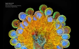
- 1. Rogelio Moreno Gill Panama, Panama Fern sorus at varying levels of maturity Fluorescence, Image Stacking
- 2. Now celebrating its 41st year, Nikon Small World is widely regarded as the leading forum to recognize proficiency and photographic excellence of photography taken under the microscope. To select the winners, competition judges analyzed entries from all over the world covering subjects ranging from chemical compounds to up-close-and-personal looks at biological specimens. (Nikon)
- 3. Dr. Joseph Parker Columbia University, Department of Genetics and Development New York, New York, USA Rove beetle head (Tychobythinus sp.)
- 4. Jose Almodovar University of Puerto Rico (UPR), Mayaguez Campus, Biology Department Mayaguez, Puerto Rico, USA Tentacles of a carnivorous plant (Drosera sp.)
- 5. Dr. Giorgio Seano & Dr. Rakesh K. Jain Harvard Medical School, Massachusetts General Hospital Edwin L. Steele Laboratory for Tumor Biology Boston, Massachusetts, USA Live imaging of perfused vasculature in a mouse bra in with glioblastoma Optical Frequency Domain Imaging System
- 6. Christian Bohley Martin Luther University Halle- Wittenberg Halle, Germany Degenerating Blue Phases (II) of 55% CB15 in E48 (s ubstance used in manufacture of Liquid Crystal Displays) Polarized Light
- 7. Dr. David Maitland Feltwell, United Kingdom Leaf cross section of a water lily leaf bud (Nuphalutea)
- 8. Douglas Moore University of Wisconsin - Stevens Point, University Relations and Communications Stevens Point, Wisconsin, USA Fairburn agate from Black Hills of western South Dakota (63x) Fiber Optic Illumination 63x
- 9. Jace Artichoker Rochester Institute of Technology (RIT) Rochester, New York, USA Mouse embryo, 10.5 days old
- 10. Norm Barker Johns Hopkins School of Medicine, Department of Pathology Baltimore, Maryland, USA Red fossil coral slab Reflected Light
- 11. Dr. Helen Rankin University of California, Berkeley Berkeley, California, USA Transgenic Xenopus laevis (African clawed toad) tadpole head expressing green neurons Confocal 10x
- 12. Dr. Igor Siwanowicz Ashburn, Virginia, USA Hughes Medical Institute (HHMI), Janelia Farm Research Campus, Leonardo Lab Intake of a humped bladderwort ( Utricularia gibba), a freshwater carnivorous plant
- 13. Donald Parsons Madison, Wisconsin, USA Scales on moth wing Image Stacking 300x
- 14. Dr. David Maitland Feltwell, United Kingdom Black witch-hazel (Trichodactylus crinitus) leaf producing crystals to defend against herbivores
- 15. Howard Lynk Morehead City, North Carolina, USA Micro-engraving on glass from an antique microscope slide created by Washington Teasdale c. 1880 Darkfield 100x
- 16. Dr. Alessio Colombo DZNE Munich, Bavaria, Germany Mouse dorsal root ganglia (neuronal plus Schwann cells) in culture Confocal
- 17. Dr. Matthew S. Lehnert Kent State University at Stark North Canton, Ohio, USA Proboscis (mouthparts) tip of a vampire moth (Calyptra thalictri).
- 18. Dr. Aleksandar Matkovic University of Belgrade, Institute of Physics Center for Solid State Physics and New Materials Belgrade, Serbia Gold and titanium electrodes covered by graphene sheet Brightfield
- 19. Dr. David Maitland Feltwell, United Kingdom Vascular bundles of papyrus (Cyperus papyrus)
- 20. Kirti Prakash Institute of Molecular Biology, Mainz Mainz, Germany DNA inside cell nucleus Super-Resolution Microscope
- 21. Dr. Igor Siwanowicz Ashburn, Virginia, USA Hughes Medical Institute (HHMI), Janelia Farm Research Campus, Leonardo Lab Antenna of a male moth (Anisota sp.)
- 22. Dr. Heiti Paves Tallinn University of Technology, Department of Gene Technology Tallinn, Estonia Anther of a flowering plant (Arabidopsis thaliana)
- 23. Michael Crutchley Pembrokeshire, Wales, United Kingdom Radula (feeding structure) of an aquatic snail (Limpet) Darkfield Epi. 40x
- 24. Dr. Luca Toledano Museo Civico di Storia Naturale di Verona Verona, Italy Detail of jewel beetle (Coleoptera Buprestidae)
- 25. Cynthia Levinthal Q Therapeutics, Clinical/Research Department Salt Lake City, Utah, USA
- 26. Frank Reiser Nassau Community College, Department of Biology Garden City, New York, USA Suction cups on the diving beetle ( Dytiscus sp.) foreleg Image Stacking, Photo- merge 50x
- 27. Charles B. Krebs Charles Krebs Photography Issaquah, Washington, USA Feeding rotifers (Floscularia ringens) Darkfield 50x
- 28. Dr. Yoji Tanaka Microphoto Studio "Cat's Glove" Ebina, Kanagawa, Japan Twinned crystals of 4,4'- dibromobiphenyl Polarized Light, Retardation Control
- 29. Dr. Maria Boulina, Dr. Akira Chiba & Hasitha Samarajeewa University of Miami Miami, Florida, USA Individually colored neurons in a live fruit fly (Drosophila) larva Fluorescence, Confocal
- 30. Dr. David Linstead Bromley, Kent, United Kingdom Buoyancy organs of a phantom midge (Chaoborus) larva Polarized Light 125x
- 31. Jacek Myslowski Wloclawek, Kujawsko-Pomorskie, Poland Alona guttata (water flea) Fluorescence 200x
- 32. Arturo Agostino Reggio Calabria, Italy Colony of single celled organisms (Carchesium ciliates) Differential Interference Contrast
- 33. Raymond Morrison Sloss Quekett Microscope Club Banbury, Oxfordshire, United Kingdom Mouth parts (pseudo trachea) of a Blowfly (Callipho ra vomitoria)
- 34. Dr. Tetsuaki Miyake York University, Department of Biology Toronto, Ontario, Canada Cultured mouse embryo cells
- 35. Harry Leung Harvard Medical School Program in Cellular and Molecular Medicine, Children's Hospital Boston Boston, Massachusetts, USA Stinger of a honey bee
- 36. Thomas Deernick University of California, San Diego La Jolla, California, USA Triple-labeled rat cerebellum Multiphoton 100x
- 37. Susan Tremblay University of California, Berkeley Berkeley, California, USA Liverwort (Lepidolaena taylorii) plant showing modified leaves (water sacs), which are often home to aquatic microorganisms such as rotifers
- 38. Daniel H. Miller & Ethan S. Sokol Whitehead Institute for Biomedical Research, Massachusetts Institute of Technology, Department of Biology Cambridge, Massachusetts, USA Lab-grown human mammary gland organoid
- 39. David Spears David Spears Imaging Taunton, Somerset, United Kingdom Root tip section of a dicot plant Differential Interference Contrast 25.5x
- 40. Dr. Nathanael Prunet California Institute of Technology and Dartmouth College, Department of Biology Pasadena, California, USA Young buds of Arabidopsis (a flowering plant)
- 41. Dr. Ariadna Recasens University of Sydney, Kolling Institute of Medical Research Sydney, Australia Micrometamorphosis: from human stem cells to neurons Fluorescence 20x
- 42. Donn Dr. Donna Beer Stolz University of Pittsburgh, Department of Cell Biology Pittsburgh, Pennsylvania, USA Mutant human alpha-1 antitrypsin aggregates (red) exiting the endoplasmic reticulum (green) in an iPS cell differentiated into a liver cell Confocal Reconstruction 200x
- 43. Katherine Pfister University of Virginia Charlottesville, Virginia, USA Xenopus laevis (frog) tadpole- ventral view
- 44. Anatoly Mikhaltsov Omsk, Russia Transverse section of an ostrich fern Brightfield 250x
- 45. Michael Crutchley Pembrokeshire, Wales, United Kingdom Adult marine worm (Autolytus) Macroscopy 30x
- 46. Jie Zh Jie Zhang University of Illinois at Urbana-Champaign Urbana, Illinois, USA Janus particles (micro- particles) suspended in water between two transparent electrode (40x) Brightfield 40x
- 47. Evan Darling Memorial Sloan Kettering Cancer Center New York, New York, USA Starfish imaged using confocal microscopy
- 48. Dr. Konstantin Bergmeister Marion Gröger, Martin Aman, Anna Willensdorfer, Krisztina Manzano- Szalai & Oskar C. Aszmann Medical University of Vienna, Christian Doppler Laboratory for Restoration of Extremity Function, Division of Plastic and Reconstructive Surgery, Vienna, Austria Murine biceps muscle stained to show different muscle fiber populations Fluorescence -immunohistochemistry
- 49. Dr. Robert Markus University of Nottingham, School of Life Sciences Super Resolution Microscopy Department Nottingham, United Kingdom Bovine pulmonary artery endothelial cells