Neglected Achilles Tendon Repair Using Flexor Hallucis Longus Augmentation
•
1 gostou•693 visualizações

IOSR Journal of Dental and Medical Sciences is one of the speciality Journal in Dental Science and Medical Science published by International Organization of Scientific Research (IOSR). The Journal publishes papers of the highest scientific merit and widest possible scope work in all areas related to medical and dental science. The Journal welcome review articles, leading medical and clinical research articles, technical notes, case reports and others.

Recomendados
Mais conteúdo relacionado
Mais procurados
Mais procurados (20)
Destaque
Destaque (20)
Semelhante a Neglected Achilles Tendon Repair Using Flexor Hallucis Longus Augmentation
Semelhante a Neglected Achilles Tendon Repair Using Flexor Hallucis Longus Augmentation (20)
Mais de iosrjce
Mais de iosrjce (20)
Último
Último (20)
Neglected Achilles Tendon Repair Using Flexor Hallucis Longus Augmentation
- 1. IOSR Journal of Dental and Medical Sciences (IOSR-JDMS) e-ISSN: 2279-0853, p-ISSN: 2279-0861.Volume 14, Issue 11 Ver. VI (Nov. 2015), PP 05-09 www.iosrjournals.org DOI: 10.9790/0853-141160509 www.iosrjournals.org 5 | Page Neglected Tendo-Achilles Rupture Repair by Fhl Augmentation Using Bio-Screw and Pull out Suture Using Suture Plastic Wastes: An Innovative Technique Dr Praveen Kumar Pandey,MS Ortho.; Dr Inder Pawar, MS Ortho.; Dr Jyoti Gupta, MD Anaesthesia; Dr Raaghav Rai Verma, MS Ortho. Abstract: Injuries of the Achilles tendon are relatively common in middle-aged athletes. Achilles tendon ruptures have been estimated to be the third most frequent tendon rupture. Most commonly, the mechanisms of Achilles tendon rupture are pushing off with the weight bearing forefoot while extending the knee, sudden unexpected dorsiflexion of the ankle and violent dorsiflexion of the plantar flexed foot, as in a fall from a height. The decision to treat acute Achilles tendon ruptures conservatively or operatively is somewhat controversial, with evidence to support either form of treatment. We have operated three cases of tendo-achilles rupture with tear very close to the insertion site over calcaneus, all three cases were chronic neglected ruptures with patients in range of 40 – 50 years of age. All the patients were subjected to clinical examination, ultrasonographic evaluation and prepared for tendon transfer. The surgical technique consists of transfer of flexor hallucis longus (FHL) tendon as described by Wapner in two patients with modification using bio-screws and direct repair of neglected rupture using V-Y advancement of gastro-soleus aponeurosis as described by Abraham and Pankovich. The Achilles tendon is exposed with hockey-shaped postero-medial direct incision. The FHL tendon is interlaced distally with krackow suturing technique using ethibond suture and fixed to the calcaneum with bio-screws. The Achilles tendon stump is passed in a conical vertical trans-calcaneal tunnel to exit out of the middle of the heel with Krackow suture using 2-0 non-absorbable suture in place, this pull-out suture with tendoachilles tendon proximal stump anchored over the heel using plastic wastes of suture material with ankle in` 5 to 10° of plantar flexion. In the third case, after doing V-Y advancement of gastro-soleus aponeurosis and putting krackow suture using 2-0 non-absorbable suture in proximal stump of tendo-achilles, this pull-out suture with tendoachilles stump anchored over heel using plastic wastes of suture material with ankle in 5 to 10° of plantar flexion. Augmented repairs provide stronger reconstruction and gives more biomechanical stability to the repair. The current approach to the rehabilitation of Achilles tendon rupture surgery is based on a short period of immobilization and early weight-bearing because of the complications due to prolonged immobilization. In conclusion, our case series of three patients, using augmentation by FHL tendon fixed to the calcaneum with bio-screw and using plastic wastes of suture material in place of button for anchorage of pull-out suture over heel due to unavailability of buttons in the operation theatre found to have good results in the form of functional outcome and easy availability of sterile material for anchorage of pull-out suture. Keywords: Achilles tendon, neglected rupture, flexor hallucis longus, V-Y advancement, bio-screw, pull-out suture, suture plastic wastes. I. Introduction Since first described by Ambroise Paré in 1575 and reported in 1633 in the literature, rupture of the Achilles tendon has received increasing attention regarding treatment.1 Injuries of the Achilles tendon are relatively common in middle-aged athletes and the frequency of these injuries is increasing as more people remain active in recreational sports and activities for longer times. Achilles tendon (TA) ruptures have been estimated to be the third most frequent tendon rupture. The peak age for TA ruptures in both men and women is between 30 and 40 years of age. Most commonly, the mechanisms of TA rupture are pushing off with the weight bearing forefoot while extending the knee, sudden unexpected dorsiflexion of the ankle and violent dorsiflexion of the plantar flexed foot, as in a fall from a height. Disruption also can occur from a direct blow to the contracted tendon or from a laceration. TA rupture has been related to a relatively hypovascular area of the tendon, shown by angiography to be 2 to 6 cms above insertion into the calcaneus. The cause of Achilles tendon rupture probably is a combination of a relatively hypovascular area and repetitive trauma that causes an inflammatory reparative process that is unable to keep up with the stresses because of decreased vascularity. A mechanical overload completes the rupture. The decision to treat acute TA ruptures conservatively or operatively is somewhat controversial, with evidence to support either form of treatment, but surgical repair seems to have been the preferred treatment in the late 1980s and 1990s.2-5 Non-operative treatment begins with immobilization of the ankle in plantar flexion for
- 2. Neglected Tendo-Achilles Rupture Repair by Fhl Augmentation Using Bio-Screw and Pull out... DOI: 10.9790/0853-141160509 www.iosrjournals.org 6 | Page 6- 8 weeks using plaster casts.6 Although non-operative treatment avoids the risk of surgery and decreases patient cost, this may result in a lengthened tendon with reduced power of the gastrosoleus muscle7,8 and a high re-rupture rates.2,4 A number of techniques have been described in the literature for reconstruction of a neglected TA rupture. If the tendon defect is less than 3 cms after debridement and the injury is less than 3months old, direct repair often is possible. If, however, the tendon gap is more than 3 cm (more common), additional techniques must be used, such as local tissue transfer, tissue augmentation, synthetics and allografts. V-Y quadriceps-plasty and gastro-soleus aponeurosis turn down flap can be used for augmentation, and local tendon transfers (flexor hallucis longus, flexor digitorum longus, peroneus brevis and plantaris longus) can be used to bridge larger defects. The purpose of this study was to report the results of our case series of chronic TA ruptures using the innovative technique of augmented repair and anchorage of pull-out sutures over heel using plastic wastes of suture material in place of routinely used buttons. II. Case Report We have operated three cases of TA rupture with tear very close to the insertion site over calcaneus(within 2 cm. of insertion) found intra-operatively, all three cases were chronic neglected ruptures with patients in range of 40 – 50 years of age. Duration from rupture to surgical treatment was 4 months to 6 months in all the three cases. Tendon gap was palpable in all the three cases while in two cases a clear gap of over 3 cms was palpated from outside and is indicative of the real gap left in the tendon after debridement and this is one of the reason we feel is a clear indication for tendon transfer in preference to direct suture. All three patients were subjected to clinical examination , Ultrasonographic evaluation and two of them prepared for tendon transfer. The surgical technique consists of transfer of flexor hallucis longus (FHL) tendon as described by Wapner in two patients with modification of using bio-screw for fixation to calcaneus and direct repair of neglected rupture using V-Y advancement of gastro-soleus aponeurosis as described by Abraham and Pankovich in one patient. The Achilles tendon is exposed with hockey-shaped postero-medial direct incision in two cases and direct posterior incision over gastro-soleus muscle and Achilles tendon in one case. The tendon morphology is studied and the ends were debrided adequately. In two cases, FHL tendon is isolated on the medial side of the foot and is retrieved through the postero-medial incision. The FHL tendon is interlaced distally with krackow suturing technique using ethibond suture and fixed to the calcaneum with bio-screws through a vertical tunnel above downwards. The Achilles tendon stump is passed in a conical vertical trans-calcaneal tunnel to exit out of the middle of the heel with Krackow suture using 2-0 non-absorbable suture in place, this pull-out suture with tendo- achilles tendon stump anchored over the heel using plastic wastes of suture material with ankle in 5 to 10° of plantar flexion. This method was also used in the third case, after doing V-Y advancement of gastro-soleus aponeurosis and putting krackow suture using 2-0 non-absorbable suture in proximal stump of tendo-achilles, this pull-out suture with tendo-achilles stump anchored over the heel using plastic wastes of suture material with ankle in 5 to 10° of plantar flexion. An above knee posterior splint is applied after insertion of suction drain. CASE 1 - 45 years old male presented to Orthopaedics out patient department (OPD) with 4 months old chronic neglected TA rupture due to sudden dorsiflexion of left feet while climbing stairs with weight on his head. Patient found to have pain in the heel and difficulty in walking with insufficient push-off during terminal stance phase on examination. Intra-operatively, tendon gap was found to be 5 cm. after debridement (figure 1) and operated with augmented repair using FHL fixed to the calcaneus with bio-screw through a vertical tunnel above downwards and pull-out suture technique as described above. CASE 2 - 49 years old male presented to Orthopaedics OPD with 5 months old chronic neglected TA rupture right due to fall from height of 10 ft. on both feet. Patient presented with complaint of difficulty in walking on uneven ground. Patient found to have insufficient push-off during terminal stance phase. Intra- operatively, tendon gap was found to be 5 cm (figure 2). after debridement and operated with augmented repair using FHL fixed to the calcaneus with bio-screw through a vertical tunnel above downwards and pull-out suture technique as described above. CASE 3 - 48 years old male presented to Orthopaedics OPD with 4 ½ months old chronic neglected TA rupture left due to sudden dorsiflexion of ankle while loading weight on his head, labourer by occupation. Patient presented with pain in the heel and difficulty in walking with palpable defect over tendo-achilles. Patient found to have insufficient push-off during terminal stance phase. Intra-operatively, tendon gap was found to be 2 cm. after debridement and operated with direct repair after V-Y advancement of gastro-soleus aponeurosis (figure 3). The repair is strengthened with pull-out suture technique as described above (figure 4 &5). The postoperative protocol consists of first dressing at 5th day and suture removal at 2 weeks, pull out suture retrieval and full weight-bearing at 6 weeks. Isokinetic & isometric strengthening and proprioceptive exercises were applied to the extremity for 3 weeks under control of an experienced physical rehabilitation team at the same time. Running on an even surface was not allowed before 12 weeks after the operation. Previous athletic activity was allowed at the sixth month. The follow-up period ranged from 1.5 to 2 years. The results
- 3. Neglected Tendo-Achilles Rupture Repair by Fhl Augmentation Using Bio-Screw and Pull out... DOI: 10.9790/0853-141160509 www.iosrjournals.org 7 | Page were evaluated by the American orthopaedic Foot and Ankle society (AOFAS) hind foot score which was found to be very good in all the three cases. III. Discussion TA rupture most commonly occurs in 25–40 years old active individuals. 90% of the patients injured the tendon while doing some form of recreational activities. Although age-related risk factors for Achilles tendon injuries are well reported in the English literature, however there is no studies that analyze the relationship between the specific sports and Achilles tendon injuries.9,10 The aetiology of the Achilles tendon rupture remains unclear, but some of the investigations have supported the theory of chronic degenerative changes based on histological examination of material obtained from the ruptured area during the operation.11,12 Inglis and Sculco proposed that there is an inhibitor mechanism that regulates the length versus power mechanism of the musculo-tendinous unit, which limits the tendon length when sudden overloading of the tendon is applied.3 Treatment options available for TA rupture, such as non-operative closed methods, open surgical repair or percutaneous sutures, which have long been a matter of controversy. The disadvantages of closed procedures are high re-rupture rates (10–30%) and less strength & endurance compared with open surgical repair.13-15 The proper indications for surgical repair appear to be an active patient who demands to return to functional status at the earliest possible with a short rehabilitation program. Meanwhile, with the developments of operation techniques, the complications of open repair have become less frequent. However, Nistor found only minor differences between the results of surgical and non-surgical treatment.13 Simple end-to-end suture is easier to perform and requires a less-extensive dissection, but to approximate a poor quality tendon with only end-to-end suture is not safe. Less-invasive techniques also have been developed to perform end-to-end suture of the Achilles tendon percutaneously.8,16 The incidence of sural nerve injury and re-rupture rate seems to be higher with percutaneous repairs.17 In cases of neglected chronic TA ruptures, augmented TA repair provides stronger reconstruction and provides more biomechanical stability to the repair. Central gastro-soleus aponeurosis flap repair is superior to standard Kessler repair by virtue of its strength. Augmentation by adding collagen to the repair site allows earlier mobility, weight-bearing and a more aggressive rehabilitation program with reduction in the incidence of re- rupture for both acute and neglected Achilles tendon rupture.18,19 In the recent past, regardless of the treatment methods – non-operative closed methods, open surgical methods or percutaneous procedures – casting in ankle equines without weight-bearing for a minimum of 6–8 weeks has been widely accepted. The current approach to the rehabilitation of TA rupture surgery is based on a short period of immobilization and early weight-bearing because of the complications due to prolonged immobilization, such as arthrofibrosis, joint stiffness, calf atrophy, damage to the articular cartilage and deep vein thrombosis.20,21 Also, prolonged immobilization and limitation of weight-bearing accompanied with prolonged rehabilitation program and sick leave time and resultant a weakened, atrophic & less vascularized tendon that is prone to re-ruptures. Although the positive effects of early mobilization on the healing process after surgical repair have been well documented by several reports,22,23 in these studies full weight-bearing was not allowed for 4–6 weeks. Tensile loads cause viscoelastic response at the muscle–tendon unit and the ruptures are most commonly seen at the musculo-tendinous junction. Early full weight-bearing after surgical repair by protection from overloading may allow newly formed collagen fibre to grow and be remodelled rapidly, ultimately sustaining good tendon strength.24,25 In the majority of the published series, the results of the primary repair have been reported about TA rupture surgery rehabilitated with early weight-bearing rather than augmented reconstruction. In our series, the clinical results of augmented repair of Achilles tendon using bio- screws and plastic wastes of suture material in place of button for anchorage of pull-out suture over heel due to unavailability of buttons in the operation theatre with early weight-bearing in walking cast demonstrated satisfactory functional recovery without any major complications. All the three cases showed good results at one year follow-up in terms of the American orthopaedic Foot and Ankle society (AOFAS) hind foot score. Surgical treatment of TA rupture is associated with a significantly lower incidence of re-rupture and therefore is the treatment method of choice. Non-surgical treatment may be acceptable for patients who refuse surgery or who are unfit for surgery. Functional early mobilisation appears to be associated with an improved functional outcome and should be considered in preference to plaster cast immobilisation where appropriate.26 Nevertheless, augmented repair of the Achilles tendon rupture has certain handicaps. The major disadvantages of augmented reconstruction are increased rate of wound complication and infection due to the more extensive approach.27 Deep infection after surgical repair of TA rupture is a relatively rare but devastating problem as the skin and soft-tissue defects around the ankle are major challenges for the surgeon. Methods for the reconstruction of soft tissue defects after infection have been reported in the literature, but the results found to be variable and insufficient data is available in general to support any of the techniques.28,29 Another problem
- 4. Neglected Tendo-Achilles Rupture Repair by Fhl Augmentation Using Bio-Screw and Pull out... DOI: 10.9790/0853-141160509 www.iosrjournals.org 8 | Page is widening of the posterior aspect of the ankle. The repaired part of the tendon heals with a bulky tissue that might results in contact ulcers with shoeing. Zell and Santoro reported no re-rupture in their augmented repair series of 25 acute TA rupture.19 Although it was reported that re-ruptures mostly result from full weight-bearing during the first few days after removal of the cast or orthosis.20,30 We did not encounter any immediate re-rupture after removal of the cast in our series due to gradual progression to full weight-bearing after cast removal. However, re-rupture is a troublesome complication that is difficult situation for both the surgeon and the patient. The sural nerve injury can also overshadow the success of the operation even if most of the injuries are transient.19,27 In order to prevent this complication, the sural nerve can be protected in the lateral skin flap of the posteromedially placed skin incision. In our series, no patient had sural nerve injuries and any other major complications. We believe that expectations of professional athletes from surgical treatment are more demanding than from non-professionals. However, all of the patients in our series demanded return to a particular level of pre- injury activity at the earliest date possible. In our series all three patients achieved pre-injury activity status with slight limitation at approximately one year follow up. In conclusion, augmented repair of chronic TA ruptures using tendon grafts are strong and stable enough to allow early weight-bearing ambulation with favourable clinical results in most of the patients. In our series, new innovative technique of using bio-screw fixation for FHL augmentation and using plastic wastes of suture material in place of button for anchorage of pull-out suture over heel due to unavailability of buttons in the operation theatre found to have good results in the form of functional outcome and easy availability of sterile material for anchorage of pull-out suture. The disadvantages of the procedure have to be shared in detail with patients before the operation. Care must be taken about keeping dressing gauge in between heel and plastic material of suture and tying the knot tightly and properly over the plastic material of suture. Further studies that include a higher number of neglected TA rupture patients need to be performed to elucidate the security and safety of this new innovated augmented technique. IV. Compliance With Ethical Standards: Funding: Not Applicable (Na). Conflict Of Interest: All the authors have no conflicts of interest. Ethical Approval: All procedures performed in our case report involving human participant were in accordance with the ethical standards of the institutional and/or national research committee and with the 1964 Helsinki declaration and its later amendments or comparable ethical standards. Informed Consent: Informed consent was obtained from the all the patients included in our study. References [1]. Popovic N, Lemaire R. Diagnosis and treatment of acute ruptures of the Achilles tendon. Current concepts review. Acta Orthop Belg. 1999;65:458–71. [2]. Edna TH. Non-operative treatment of Achilles tendon ruptures. Acta Orthop Scand. 1980;51:991–3. [3]. Inglis AE, Sculco TP. Surgical repair of ruptures of the tendo Achillis. Clin Orthop. 1981;156:160–9. [4]. Wills CA, Washburn S, Caiozzo V, Prietto CA. Achilles tendon rupture. A review of the literature comparing surgical versus nonsurgical treatment. Clin Orthop Relat Res. 1986;207:156–63. [5]. Beskin JL, Sanders RA, Hunter SC, Hughston JC. Surgical repair of Achilles tendon ruptures. Am J Sports Med. 1987;15:1–8. [6]. Cetti R, Cristiansen S-E, Ejsted R, Jensen NM, Jorgensen U. Operative versus nonoperative treatment of Achilles tendon rupture: A prospective randomized study and review of the literature. Am J Sports Med. 1993;21:791–9. [7]. Inglis AE, Scott WN, Sculco TP, Patterson AH. Ruptures of the Tendo Achilles: An objective assessment of surgical and non surgical treatment. J Bone Joint Surg Am. 1976;58:990–3. [8]. Lecestre P, Germanville T, Delplace J. and the Société Orthopédique Rochelaise. Achilles tendon ruptures treated by percutaneous tenorraphy: Multicentric study of 60 cases. Eur J Orthop Surg Traumatol. 1997;7:37–40. [9]. Kannus P, Natri A. Etiology and pathophysiology of tendon ruptures in sports. Scand J Med Sci Sports. 1997;7:107–12. [10]. Jarvinen TA, Kannus P, Maffulli N, Khan KM. Achilles tendon disorders: Etiology and epidemiology. Foot Ankle Clin. 2005;10:255–66. [11]. Barfred T. Experimental rupture of the Achilles tendons. Acta Orthop Scand. 1971;42:406. [12]. Kannus P, Jozsa L. Histopathological changes preceding spontaneous rupture of a tendon. J Bone Joint Surg Am. 1991;73:1507–25. [13]. Nistor L. Surgical and non-surgical treatment of Achilles tendon ruptures. A prospective randomized study. J Bone Joint Surg Am. 1981;63:394–9. [14]. Shields CL, Jr, Kerlan RK, Jobe FW, Carter VS, Lombardo SJ. The Cybex II evaluation of surgically repaired Achilles tendon ruptures. Am J Sports Med. 1978;6:369–72. [15]. Ma GW, Griffith TG. Percutaneous repair of acute closed ruptured Achilles tendon. A new technique. Clin Orthop Relat Res. 1997;128:247–55. [16]. Vivaldi AS. Percutaneous repair of ruptured tendo Achilles. J Bone joint Surg Br. 2000;82:619. [17]. Bradley JP, Tibone JE. Percutaneous and open surgical repair of the Achilles tendon rupture. Am J Sports Med. 1990;18:188–95. [18]. Takao M, Ochi M, Naito K, Uchio Y, Matsusaki M, Oae K. Repair of neglected Achilles Tendon rupture using gastrosoleus fascial falps. Arch Orhtop Trauma Surg. 2003;123:471–4. [19]. Zell RA, Santoro VM. Augmented repair of acute Achilles tendon ruptures. Foot Ankle Int. 2000;21:469–74.
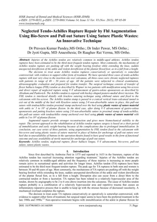
- 5. Neglected Tendo-Achilles Rupture Repair by Fhl Augmentation Using Bio-Screw and Pull out... DOI: 10.9790/0853-141160509 www.iosrjournals.org 9 | Page [20]. Aoki M, Ogiwara N, Ohta T, Nabeta Y. Early active motion and weightbearing after cross-stitch achilles tendon repair. Am J Sports Med. 1998;26:794–800. [21]. Kangas J, Pajala A, Siira P, Hämäläinen M, Leppilahti J. Early functional treatment versus early immobilization in tension of the musculotendinous unit after Achilles rupture repair: A prospective, randomized, clinical study. J Trauma. 2003;54:1171–80. [22]. Sölveborn SA, Moberg A. Immediate free ankle motion after surgical repair of acute Achilles tendon ruptures. Am J Sports Med. 1994;22:607–10. [23]. Mandelbaum BR, Mayerson MS, Foster R. Achilles tendon ruptures: A new method of repair, early range of motion, and functional rehabilitation. Am J Sports Med. 1995;23:392-5. [24]. Kubota H, Manske PR, Aoki M, Pruitt DL, Larson BJ. Effects of motion and tension on injured flexor tendons in chickens. J Hand Surg. 1996;21:456–63. [25]. Mortensen NH, Skov O, Jensen PE. Early motion of the ankle after operative treatment of a rupture of the Achilles tendon. A prospective, randomized clinical and radiographic study. J Bone Joint Surg Am. 1999;81:983–90. [26]. 26.Achilles tendon rupture: surgical versus non-surgical treatment. Lynch RM1 . Accid Emerg Nurs. 2004 Jul;12(3):149-58. [27]. Garabito A, Miranda JM, Sotelo JS. Augmented repair of acute Achilles tendon ruptures using gastrosoleus-soleus fasia. Int Orthop. 2005;29:42–6. [28]. Leppilahti J, Kaarela O, Teerikangas H, Raatikainen T, Orava S, Waris T. Free tissue coverage of wound complications following Achilles tendon rupture surgery. Clin Orthop Relat Res. 1996;328:171–6. [29]. Ademoglu Y, Ozerkan F, Ada S, Bora A, Kaplan I, Kayalar M, et al. Reconstruction of skin and tendon defects from wound complications after Achilles tendon rupture. J Foot Ankle Surg. 2001;40:158–65. [30]. Marti RK, van der Werken C, Schutte PR, Bast TJ. Operative repair of ruptured achilles tendon and functional after-treatment-I. Acute rupture. Neth J Surg. 1983;35:61–4. Figure 1 figure 2 figure 3 Figure 4 figure 5 Legends of figures: Figure 1- Intraoperative picture of case 1 showing TA rupture with tendon gap. Figure 2- Intraoperative picture of case 2 showing TA rupture with tendon gap. Figure 3- intraoperative picture of case 3 with V-Y plasty. Figure 4- clinical picture showing pull out suture anchored over heel using plastic wastes of suture material. Figure 5- clinical picture of case 1 after closure.