
Recomendados
Recomendados
Mais conteúdo relacionado
Mais procurados
Mais procurados (20)
Semelhante a Phototoxicity in live cell imaging workshop CYTO2018
Semelhante a Phototoxicity in live cell imaging workshop CYTO2018 (20)
Último
Último (20)
Phototoxicity in live cell imaging workshop CYTO2018
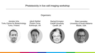
- 1. Jaroslav Icha Turku Centre for Biotechnology Turku, Finland Phototoxicity in live cell imaging workshop Jakub Nedbal Photon Force Edinburgh, UK Silas Leavesley University of South Alabama Mobile, USA Organizers: Rachel Errington Cardiff University Cardiff, UK
- 2. The workshop will be recorded We record audio for the sole purpose of note taking and it will not be published anywhere. The slides will be available. Workshop report will appear as a part of Cytometry Part A manuscript towards the end of 2018.
- 3. 1. Introductory talk 25 min: current understanding of the problem of phototoxicity and questions related directly to the talk. 2. Collecting personal experience with phototoxicity from our expert panel and anyone else who would like to contribute. 3. Sli.do online questionnaire. 4. Defining realistic guidelines or SOP for live fluorescence microscopy experiments: • controls to evaluate phototoxicity • strategies to mitigate phototoxicity • what is the biggest barrier to adoption of effective strategies to limit phototoxicity? • is there enough incentive for scientists to ensure phototoxicity is not affecting their data? • Should guidelines such as “minimal information for publication of live fluorescence imaging experiments” exist and be demanded by journals before publication? 5. Collecting input for the workshop report in Cytometry Part A. 6. Group photo. Workshop agenda
- 4. Phototoxicity in live imaging and how to avoid it Jaroslav Icha CYTO 2018 Workshop
- 5. Compromising is inevitable in live imaging experiments The photon budget concept Health of the sample must have priority over resolution. Laissue et al., 2017 Nat MethodsLiu et al., 2015, Molecular Cell
- 6. Two principal sources of phototoxicity • Destruction of endogenous molecules: Absorption in the visible light spectrum used for excitation. (violet and blue light much worse than red and farred) • ROS generated by photobleaching of fluorophores introduced to the sample (photosensitization): Can be the dominant source of phototoxicity.
- 7. Fluorophore photobleaching Magidson&Khodjakov, 2013 Methods in Cell Biol FluorescenceExcitation +O2>>> bleaching + ROS production
- 8. Photosensitization by fluorophores KillerRed: tunnel for oxygen right into the β barrel towards the fluorophore. Magidson&Khodjakov, 2013 Methods in Cell Biol FluorescenceExcitation Carpentier et al., 2009 FEBS letters +O2>>> bleaching + ROS production
- 9. Photobleaching is not a reliable readout for phototoxicity Magidson&Khodjakov, 2013 Methods in Cell BiolCentrin-GFP, HeLa cells, wide-field Higher intensity illumination
- 10. Photobleaching is not a reliable readout for phototoxicity Magidson&Khodjakov, 2013 Methods in Cell BiolCentrin-GFP, HeLa cells, wide-field Higher intensity illumination Lower intensity illumination
- 11. Icha, Weber et al., 2017, Bioessays Sample morphology is not a reliable readout for phototoxicity Overlooking phototoxicity in your experiment can be easy
- 12. Jemielita et al., 2014, Journal of Biophotonics Subtle phototoxicity resulting in qualitative changes in the samples Zebrafish craniofacial bone morphogenesis Confocal SPIM
- 13. Jemielita et al., 2014, Journal of Biophotonics Subtle phototoxicity resulting in qualitative changes in the samples Tinevez et al., 2017, Methods Zebrafish craniofacial bone morphogenesis Cumulativedistribution ParticlesofproteinNEMO Confocal SPIM Change from random walk to directionally persistent movement
- 14. Icha, Weber et al., 2017, Bioessays Detecting and quantifying phototoxicity through dose-response curves Phototoxicity does not scale linearly with the illumination intensity.
- 15. Tinevez et al., 2012, Methods Enzymol Icha, Weber et al., 2017, Bioessays Detecting and quantifying phototoxicity through dose-response curves Schmidt et al., 2017, BioRxiv Phototoxicity does not scale linearly with the illumination intensity. C.elegans embryo cell divisions. S. cerevisiae cell cycle.
- 16. What can we do about phototoxicity?
- 17. Media hacks: • Removing vitamin B2 riboflavin • Imaging at 2% O2 + Trolox/TQ • Adding ascorbate Imaging media improvements Tsunoyama et al., 2018, Nat Chem Biol
- 18. Media hacks: • Removing vitamin B2 riboflavin • Imaging at 2% O2 + Trolox/TQ • Adding ascorbate Pulsed illumination to give fluorophores time to relax back to the ground state Tsunoyama et al., 2018, Nat Chem Biol Icha, Weber et al., 2017, Bioessays
- 19. The rate of delivery of light to the sample matters. Eric Betzig: “This suggests that the instantaneous peak power delivered to the specimen may be an even more important metric of cell health than the total photon dose and should enable extended 3D observation of endogenous levels of even sparsely expressed proteins produced by genome editing.” Longer exposure time lowers phototoxicity Mubaid&Brown, 2017, Microscopy Today Paxillin-GFP,CHOcells,Wide-field
- 20. The rate of delivery of light to the sample matters. Eric Betzig: “This suggests that the instantaneous peak power delivered to the specimen may be an even more important metric of cell health than the total photon dose and should enable extended 3D observation of endogenous levels of even sparsely expressed proteins produced by genome editing.” Longer exposure time lowers phototoxicity Mubaid&Brown, 2017, Microscopy Today Liu et al., 2015, Molecular Cell Supralinear dependency of peak intensity and sample damage Paxillin-GFP,CHOcells,Wide-field
- 21. Super resolution microscopy and phototoxicity Shroff et al., 2008, Nat Biotechnology Live cell superresolution imaging requires extra careful experimental planning • Measuring cell edge dynamics with DIC • TIRF illumination • Using more light resistant cell lines (CHO, NIH3T3 cells) Focal adhesions tdEos-paxillin NIH 3T3 cells
- 22. Super resolution microscopy and phototoxicity Shroff et al., 2008, Nat Biotechnology Liu et al., 2015, Dev Cell Live cell superresolution imaging requires extra careful experimental planning • Measuring cell edge dynamics with DIC • TIRF illumination • Using more light resistant cell lines (CHO, NIH3T3 cells) Focal adhesions tdEos-paxillin NIH 3T3 cells
- 23. Alternative illumination strategies to reduce phototoxicity Liu et al., 2015, Molecular Cell
- 24. The thicker the specimen, the bigger is the advantage of restricting illumination to the focal plane in 3D imaging. Gentle imaging through selective illumination of the focal plane Icha, Weber et al., 2017, Bioessays MicroscopyU Light sheet microscopy page
- 25. Spinning disk confocal vs. Light sheet microscopes (SPIM) Light sheet illumination makes a difference Microtubule plus tips (EB3-GFP) in HUVEC cells Wu et al., 2013, Nature Biotechnology
- 26. Spinning disk confocal vs. Light sheet microscopes (SPIM) Direct comparisons between microscopes Saias et al., 2015, Dev Cell Microtubule plus tips (EB3-GFP) in HUVEC cells Wu et al., 2013, Nature Biotechnology Drosophila dorsal closure, E-cadherin-GFP
- 27. Leung et al., 2011, Development Icha, Weber et al., 2017, Bioessays Direct comparisons between microscopes Phototoxicity in the spinning disk confocal revealed by cell cycle lengthening.
- 28. Optimized detection can allow reduced illumination Icha, Weber et al., 2017, Bioessays mCherry fluorescence emission spectrum
- 29. Optimized detection can allow reduced illumination Icha, Weber et al., 2017, Bioessays
- 30. Optimized detection can allow reduced illumination Icha, Weber et al., 2017, Bioessays
- 31. Wu et al., 2013 Nat Biotech Generating quantifiable, not necessarily pretty data C. Elegans embryonic development from 4-cell stage, GFP-histone transgenic line
- 32. Denoising algorithms: allow to dramatically reduce the illumination levels. Carlton et al., 2010, PNAS Computational methods: to extract more from less Cell nuclei (magenta) X chromosome (green) Drosophila larva
- 33. Denoising algorithms: allow to dramatically reduce the illumination levels. Deep learning: 10-60× less illumination to get the same SNR, reconstruction of undersampled images in axial direction, resolution improvement… Weigert et al., 2018, bioRxiv Carlton et al., 2010, PNAS Computational methods: to extract more from less Cell nuclei (magenta) X chromosome (green) Drosophila larva Cell nuclei Schmidtea mediterranea (Planarian)
- 34. Computational methods: to extract more from less Content-aware image restoration (CARE) Neuronal network trained on synthetic images Weigert et al., 2018, bioRxiv
- 35. 1. Be aware of the problem of subtle phototoxicity. 2. Absence of cell death or obvious photobleaching is not a proof of safe illumination levels. 3. For a new experimental setup measure the phototoxicity (dose-response) curves. 4. Beware of additive effects of drugs, mutations that could sensitize the sample. 5. Optimize illumination: use light sheet, longer exposure time with lower peak intensity, longer excitation wavelength, SNR only as high as required. 6. Optimize detection: refractive index matching, minimized background, optimized filters, sCMOS cameras. 7. Use transmitted light channel to check the sample health during and at the end of the experiment. 8. Compare imaged samples to non-imaged controls. 9. Compare the results to an alternative experiment not based on live imaging. 10.Report in your publications the issues with phototoxicity and the details of your imaging settings. >>> High quality, reproducible dataset, which can serve as a basis for quantitative statements Summary
- 37. jaroslav.icha@utu.fi • Caren Norden and Norden lab members • Johanna Ivaska and Ivaska lab members • Michael Weber, HMS Cell biology microscopy facility • Christopher Schmied, Tomancak lab, MPI-CBG • Light microscopy facility of MPI-CBG Turku Collegium for Science and Medicine @IchaJaroslav Thank you Funding: Contact:
- 39. Workshop agenda 1. Introductory talk 25 min: current understanding of the problem of phototoxicity and questions related directly to the talk. 2. Collecting personal experience with phototoxicity from our expert panel and anyone else who would like to contribute. 3. Sli.do online questionnaire. 4. Defining reasonable guidelines or SOP for live fluorescence microscopy experiments: • controls to evaluate phototoxicity • strategies to mitigate phototoxicity • what is the biggest barrier to adoption of effective strategies to limit phototoxicity? • is there enough incentive for scientists to ensure phototoxicity is not affecting their data? • Should guidelines such as “minimal information for publication of live fluorescence imaging experiments” exist and be demanded by journals before publication? 5. Collecting input for the workshop report in Cytometry Part A. 6. Group photo.
- 40. Slido – Polling system
- 41. • transmitted light to monitor the health of the sample during imaging. • record a non-illuminated control region of the sample only with transmitted light in parallel to your experiment. • running the dose-response curve experiment for new experimental setups • check for changes in cell cycle (mitotic index), occurrence of membrane blebbing, delayed hatching of embryos, etc. between experimental sample and non-imaged control. • monitor viability of the samples after the imaging experiment is completed, e.g., if cells will undergo mitosis, or embryos will keep developing. • if you use imaging techniques that require high energy input (confocal, super-resolution, etc.), think about repeating the experiment at different illumination light levels or with a less intrusive setup (TIRF, light sheet, wide-field) as an additional control. Controls for phototoxicity
- 42. • Media composition: antioxidants, low oxygen, removing riboflavin • Illumination pattern changes: longer wavelength and exposure times, pulsed illumination • Selective illumination: light sheet, TIRF, HILO, two photon • Image restoration algorithms: denoising, artificial neuronal networks Strategies to mitigate phototoxicity
- 43. Workshop agenda 1. Introductory talk 25 min: current understanding of the problem of phototoxicity and questions related directly to the talk. 2. Collecting personal experience with phototoxicity from our expert panel and anyone else who would like to contribute. 3. Slido online questionnaire. 4. Defining reasonable guidelines or SOP for live fluorescence microscopy experiments: • controls to evaluate phototoxicity • strategies to mitigate phototoxicity • what is the biggest barrier to adoption of effective strategies to limit phototoxicity? • is there enough incentive for scientists to ensure phototoxicity is not affecting their data? • Should guidelines such as “minimal information for publication of live fluorescence imaging experiments” exist and be demanded by journals before publication? 5. Collecting input for the workshop report in Cytometry Part A. 6. Group photo.