
Recommended
More Related Content
What's hot
What's hot (20)
Similar to Pupillary reflexes physiology
Similar to Pupillary reflexes physiology (20)
Recently uploaded
Recently uploaded (20)
Pupillary reflexes physiology
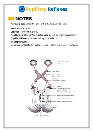
- 1. Pupillary Reflexes Normal pupil: Limits the amount of light reaching retina. Number: one pupil Location: in the center iris. Pupillary constrictor/ sphincter-innervated by parasympathetic Pupillary dilator – innervated by sympathetic visual pathway: visual center located in occipital lobe (within the calcarine sulcus).
- 2. Pupillary Reflexes Light reflex: - Pupillary light reflex (PLR) : that controls the diameter of the pupil, in response to the intensity of light that falls on the retinal ganglion cells of the retina in the back of the eye. - Stimulus: direct light (torch). - Affrent: optic nerve fibers(CNII). - Center: B/S - Eefrent: short ciliary nerve fibers (CNIII) - Effector organ: Pupillary constrictor muscle. - General pathway (important) Ganglion cell → optic nerve → pretectal nuclei→ Edinger-Westphal nucleus→ the ciliary ganglion→ short ciliary nerve fiber→ Pupillary constrictor.
- 3. Pupillary Reflexes - Light reflex pathway: (Further Reading) The afferent pupillary pathway originate in the retina , the axons of retinal ganglion cells pass into the optic nerve and decussate in the chiasm and pass with the optic tract to the midbrain, the pupillary fibers do not synapse with the visual fibers in the lateral geniculate body(LGB)but pass to the pretectal nuclei (PTN) at the level of the superior colliculus with intercalated fibers that pass as the efferent pupillary pathway to the Edinger-Westphal nucleus (EWN)of the oculomotor nerve (CN III)on each side (both sides) Preganglionic parasympathetic fibers run in the oculomotor nerve as it leaves the brain stem. The fibers pass downward to lie inferiorly in the inferior division of the third nerve as it enters the orbit. These fibers synapse in the ciliary ganglion and give rise to postganglionic parasympathetic myelinated short ciliary nerves. Normal response: Contraction of both eyes: stimulated eye (direct) another eye (indirect or consensual. Abnormal response: Relative afferent pupillary defect (RAPD) or Marcus Gunn pupil - shone on affected eye: normal + affected eye still dilated (no response) - shone on normal eye: normal + affected will contract together.
- 4. Pupillary Reflexes Corneal reflex: - is an involuntary blinking of the eyelids elicited by stimulation of the cornea (such as by touching with cotton or by a foreign body), though could result from any peripheral stimulus. - Stimulus: touch by cotton. - Stimulation should elicit both a direct and consensual response. - Significance: protect the eyes from foreign bodies and bright lights. - Responsible nerve: 5th cranial nerve (trigeminal nerve) - Afferent arc is mediated by the nasociliary branch of the ophthalmic branch (Vi) of the trigeminal. - Center: in the pons of brain stem - Efferent arc is the seventh (facial) nerve mainly temporal branch. - Effector organ: orbicularis oculi - Procedure: take a wisp of cotton then ask the patient to look straight ahead. Gently, brush the cotton against sclera or cornea, this will make the patient blink. - An abnormal corneal reflex may indicate either fifth nerve afferent disease or seventh nerve efferent disease or tolerance.
- 5. Pupillary Reflexes Accommodation reflex: - It is focusing mechanism. - is a reflex action of the eye, in response to focusing on a near object, then looking at distant object. - The reflex, controlled by the parasympathetic nervous system, involves three responses: 1. pupil constriction. 2. Increase biconvexity of lens. 3. Convergence (by medial rectus). - Stimulus: near object. - Center: Edinger–Westphal nucleus - Affrent: optic nerve - Efferent: both sympathetic and parasympathetic. - Effector organs: 1. the ciliary muscle, the medial rectus muscle (oculomotor nerve) 2. sphincter pupillae muscle (parasympathetic fibres).
- 6. Pupillary Reflexes Pathway: Some of the common diseases cause abnormal accommodation reflex may include: 1- Holmes–Adie syndrome: is a neurological disorder characterized by a tonically dilated pupil that reacts slowly to light but shows a more definite response to accommodation (i.e., light-near dissociation)
- 7. Pupillary Reflexes 2- Argyll Robertson pupils: - are bilateral small pupils that reduce in size on a near object (i.e., they accommodate), but do not constrict when exposed to bright light (i.e., they do not react to light). - They are a highly specific sign of neurosyphilis. 3- Horner's syndrome: is a combination of symptoms that arises when a group of nerves known as the sympathetic trunk is damaged. It is characterized by: miosis (a constricted pupil), partial ptosis (a weak, droopy eyelid), anhidrosis (decreased sweating) Done by: Ashaq Alqhtani & Waleed Aldosari