SlideShare uma empresa Scribd logo
1 de 18
Baixar para ler offline
Lecture Notes on Cardiovascular System 
Prepared By: Mark Fredderick R Abejo R.N, MAN 
MEDICAL AND SURGICAL NURSING 
Cardiovascular System 
Lecturer: Mark Fredderick R. Abejo RN, MAN 
Anatomy and Physiology of the Heart 
Cardiovascular system consists of the heart, arteries, 
veins & capillaries. The major function are circulation of blood, 
delivery of O2 & other nutrients to the tissues of the body & 
removal of CO2 & other cellular products metabolism 
Heart 
 Muscular pumping organ that propel blood into the arerial 
system & receive blood from the venous system of the body. 
 Hollow muscular behind the sternum and between the lungs 
 Located on the middle of mediastinum 
 Resemble like a close fist 
 Weighs approximately 300 – 400 grams 
 Has heart wall has 3 layers 
 Endocardium – lines the inner chambers of the 
heart, valves, chordate tendinae and papillary 
muscles. 
 Myocardium – muscular layer, middle layer, 
responsible for the major pumping action of the 
ventricles. 
 Epicardium – thin covering(mesothelium), 
covers the outer surface of the heart 
 Pericardium – invaginated sac 
 Visceral – attached to the exterior of 
myocardium 
 Parietal – attached to the great vessels and 
diaphragm 
 Papillary Muscle 
Arise from the endocardial & myocardial surface of the 
ventricles & attach to the chordae tendinae 
 Chordae Tendinae 
Attach to the tricuspid & mitral valves & prevent eversion 
during systole 
 Separated into 2 pumps: 
 right heart – pumps blood through the lungs 
 left heart – pumps blood through the peripheral 
organs 
 Chamber of the Heart 
Atria 
 2 chambers, function as receiving chambers, lies 
above the ventricles 
 
Medical and Surgical Nursing 1 Abejo
Lecture Notes on Cardiovascular System 
Prepared By: Mark Fredderick R Abejo R.N, MAN 
 Upper Chamber (connecting or receiving) 
 Right Atrium: receives systemic venous blood 
through the superior vena cava, inferior vena cava & 
coronary sinus 
 Left Atrium: receives oxygenated blood returning to 
the heart from the lungs trough the pulmonary veins 
Ventricles 
 2 thick-walled chambers; major responsibility for 
forcing blood out of the heart; lie below the atria 
 Lower Chamber (contracting or pumping) 
 Right Ventricle: contracts & propels deoxygenated 
blood into pulmonary circulation via the aorta 
during ventricular systole; Right atrium has 
decreased pressure which is 60 – 80 mmHg 
 Left Ventricle: propels blood into the systemic 
circulation via aortaduring ventricular systole; Left 
ventricle has increased pressure which is 120 – 180 
mmHg in order to propel blood to the systemic 
circulation 
 Heart Valves 
 Tricuspid 
 Pulmonic 
 Mitral 
 Aortic 
 Coronary artery – 1st branch of aorta 
Right Coronary 
 SA nodal Branch – supplies SA node 
 Right marginal Branch – supplies the right border 
of the heart 
 AV nodal branch – supplies the AV node 
 Posterior interventricular artery – supplies both 
ventricles 
Left Coronary 
 Circumflex branch – supplies SA node in 40 % of 
people 
 Left marginal – supplies the left ventricle 
 Anterior interventricular branch aka Left anterior 
descending(LAD)–supplies both ventricles and 
interventricular septum 
 Lateral branch – terminates in ant surface of the 
heart 
 Coronary Veins 
Coronary sinus – main vein of the heart 
Great Cardiac vein – main tributary of the coronary sinus 
Oblique vein – remnant of SVC, small unsignificant 
Heart Circulation 
Cardiac Conduction System 
Properties of Heart Conduction System 
• Automaticity 
• Excitability 
• Conductivity 
• Contractility 
Structure of Heart Conduction System 
 Nodal tissues 
SA Node( Sino-atrial, Keith and Flack) 
 Primary Pacemaker 
 Between SVC and RA 
 Vagal and symphatetic innervation 
 Sinus Rhythms 
Medical and Surgical Nursing 2 Abejo
Lecture Notes on Cardiovascular System 
Prepared By: Mark Fredderick R Abejo R.N, MAN 
AV Node( Atrioventricular , Kent and Tawara) 
 At the right atrium 
 3 zones 
 AN Zone(atrionodal) 
 N Zone (nodal) 
 NH zone (nodal –HIS) 
 Internodal and Interatrial Pathways 
Connects SA and AV Node 
Ant. Internodal(bachman) tract 
Middle Internodal(wenkebach) tract 
Posterior internodal(Thorel) tract 
 Bundle of His/ Purkinje Fibers 
Provides for ventricular conduction system 
Fastest conduction among cardiac tissues 
Right bundle 
Left Bundle 
Cardiac Action Potential 
 Depolarization: electrical activation of a cell caused by 
the influx of sodium into the cell while potassium exits 
the cell 
 Repolarization: return of the cell to the resting state 
caused by re-entry of potassium into the cell while 
sodium exits 
 Refractory periods: 
Effective refractory period: phase in which cells 
are incapable of depolarizing 
Relative refractory period: phase in which cells 
require a stronger-than-normal stimulus to 
depolarize 
Anatomical Sequence of Excitation of the Heart 
 (right atrium) 
 sinoatrial node (SA) 
 (right AV valve) 
 atrioventricular node (AV) 
 atrioventricular bundle (bundle of His) 
 right & left bundle of His branches 
 Purkinje fibers of ventricular walls 
(from SA through complete heart contraction = 220 ms = 0.22 s) 
a. Sinoatrial node (SA node) "the pacemaker" - has the 
fastest autorhythmic rate (70-80 per minute), and sets the 
pace for the entire heart; this rhythm is called the sinus 
rhythm; located in right atrial wall, just inferior to the 
superior vena cava 
b. Atrioventricular node (AV node) - impulses pass from 
SA via gap junctions in about 40 ms.; impulses are 
delayed about 100 ms to allow completion of the 
contraction of both atria; located just above tricuspid 
valve (between right atrium & ventricle) 
c. Atrioventricular bundle (bundle of His) - in the 
interATRIAL septum (connects L and R atria) 
d. L and R bundle of His branches - within the 
interVENTRICULAR septum (between L and R 
ventricles) 
e. Purkinje fibers - within the lateral walls of both the L and 
R ventricles; since left ventricle much larger, Purkinjes 
more elaborate here; Purkinje fibers innervate “papillary 
muscles” before ventricle walls so AV can valves prevent 
backflow 
The Normal Cardiac Cycle 
General Concepts 
Systole - period of chamber contraction 
Diastole - period of chamber relaxation 
Cardiac cycle - all events of systole and diastole during one 
heart flow cycle 
Events of Cardiac Cycle 
1. mid-to-late ventricular diastole: ventricles filled 
 the AV valves are open 
 pressure: LOW in chambers; HIGH in 
aorta/pulmonary trunk 
 aortic/pulmonary semilunar valves CLOSED 
 blood flows from vena cavas/pulmonary vein INTO 
atria 
 blood flows through AV valves INTO ventricles 
(70%) 
2. ventricular systole: blood ejected from heart 
 filled ventricles begin to contract, AV valves 
CLOSE 
 contraction of closed ventricles increases pressure 
 ventricular ejection phase - blood forced out 
 semilunar valves open, blood -> aorta & pulmonary 
trunk 
3. isovolumetric relaxation: early ventricular diastole 
 ventricles relax, ventricular pressure becomes LOW 
 semilunar valves close, aorta & pulmonary trunk 
backflow 
TOTAL CARDIAC CYCLE TIME = 0.8 second 
(normal 70 beats/minute) 
atrial systole (contraction) = 0.1 second 
ventricular systole (contraction) = 0.3 second 
quiescent period (relaxation) = 0.4 second 
Cardiac Output - Blood Pumping of the Heart 
General Concepts 
• Stroke volume: the amount of blood ejected with each 
heartbeat 
• Cardiac output: amount of blood pumped by the 
ventricle in liters per minute 
• Preload: degree of stretch of the cardiac muscle fibers at 
the end of diastole 
• Contractility: ability of the cardiac muscle to shorten in 
response to an electrical impulse 
• Afterload: the resistance to ejection of blood from the 
ventricle 
• Ejection fraction: the percent of end-diastolic volume 
ejected with each heartbeat 
Medical and Surgical Nursing 3 Abejo
Lecture Notes on Cardiovascular System 
Prepared By: Mark Fredderick R Abejo R.N, MAN 
General Variables of Cardiac Output 
1. Cardiac Output (CO) - blood amount pumped per minute 
 CO (ml/min) = HR (beats/min) X SV (ml/beat) 
 Normal CO = 75 beats/min X 70 ml/beat 
= 5.25 L/min 
2. Heart Rate (HR) - cardiac cycles per minute 
 Normal range is 60-100 beats per minute 
 Tachycardia is greater than 100 bpm 
 Bradycardia is less than 60 bpm 
 Sympathetic system INCREASES HR 
 Parasympathetic system (Vagus) DECREASES HR 
3. Blood pressure - Cardiac output X peripheral resistance 
 Control is neural (central and peripheral) and 
hormonal 
 Baroreceptors in the carotid and aorta 
 Hormones- ADH, aldosterone, epinephrine can 
increase BP; ANF can decrease BP 
Regulation of Stroke Volume (SV) 
 End diastolic volume (EDV) - total blood collected in 
ventricle at end of diastole; determined by length of 
diastole and venous pressure (~ 120 ml) 
 End systolic volume (ESV) - blood left over in ventricle 
at end of contraction (not pumped out); determined by 
force of ventricle contraction and arterial blood pressure 
(~50 ml) 
SV (ml/beat) = EDV (ml/beat) - ESV (ml/beat) 
Normal SV = 120 ml/beat - 50 ml/beat = 70 ml/beat 
Frank-Starling Law of the Heart - critical factor for stroke 
volume is "degree of stretch of cardiac muscle cells"; 
more stretch = more contraction force 
increased EDV = more contraction force 
slow heart rate = more time to fill 
exercise = more venous blood return 
Regulation of Heart Rate (Autonomic, Chemical, Other) 
1. Autonomic Regulation of Heart Rate (HR) 
 Sympathetic - NOREPINEPHRINE (NE) increases heart 
rate (maintains stroke volume which leads to increased 
Cardiac Output) 
 Parasympathetic - ACETYLCHOLINE (ACh) decreases 
heart rate 
 Vagal tone - parasympathetic inhibition of inherent rate 
of SA node, allowing normal HR 
 Baroreceptors, pressoreceptors - monitor changes in 
blood pressure and allow reflex activity with the 
autonomic nervous system 
2. Hormonal and Chemical Regulation of Heart Rate (HR) 
 epinephrine - hormone released by adrenal medulla 
during stress; increases heart rate 
 thyroxine - hormone released by thyroid; increases heart 
rate in large quantities; amplifies effect of epinephrine 
 Ca++, K+, and Na+ levels very important; 
hyperkalemia - increased K+ level; KCl used to 
stop heart on lethal injection 
hypokalemia - lower K+ levels; leads to 
abnormal heart rate rhythms 
hypocalcemia - depresses heart function 
hypercalcemia - increases contraction phase 
hypernatremia - HIGH Na+ concentration; can 
block Na+ transport & muscle contraction 
3. Other Factors Effecting Heart Rate (HR) 
normal heart rate - fetus 140 - 160 beats/minute 
female 72 - 80 beats/minute 
male 64 - 72 beats/minute 
1. exercise - lowers resting heart rate (40-60) 
2. heat - increases heart rate significantly 
3. cold - decreases heart rate significantly 
4. tachycardia - HIGHER than normal resting heart rate 
(over 100); may lead to fibrillation 
5. bradycardia - LOWER than normal resting heart rate 
(below 60); parasympathetic drug side effects; physical 
conditioning; sign of pathology in non-healthy patient 
Vascular System 
Major function of the blood vessels isto supply the tissue 
with blood, remove wastes, & carry unoxygenated blood 
back to the heart 
Types of Blood Vessels 
Arteries 
Elastic-walled vessels that can stretch during systole & 
recoil during diastole; they carry blood away from the 
heart & distribute oxygenated blood throughout the body 
Arterioles 
Small arteries that distribute blood to the capillaries & 
function in controlling systemic vascular resistance & 
therefore arterial pressure 
Capilliaries 
The following exchanges occurs in the capilliaries 
O2 & CO2 
Solutes between the blood & tissue 
Fluid volume transfer between the plasma & 
interstitial space 
Venules 
Small veins that receive blood from capillaries & 
function as collecting channels between the capillaries & 
veins 
Veins 
Low-pressure vessels with thin small & less muscles than 
arteries; most contains valves that prevent retrograde 
blood flow; they carry deoxygenated blood back to the 
heart. When the skeletal surrounding veins contract, the 
veins are compressed, promoting movement of blood 
back to the heart. 
Medical and Surgical Nursing 4 Abejo
Lecture Notes on Cardiovascular System 
Prepared By: Mark Fredderick R Abejo R.N, MAN 
Assessment of the Client with Cardiovascular 
Disorders 
Nursing History 
Risk Factors 
A. Non – Modifiable Risk Factor 
 Age 
 Gender 
 Race 
 Heredity 
B. Modifiable Risk Factor 
 Stress 
 Diet 
 Exercise 
 Sedentary lifestyle 
 Cigarette smoking 
 Alcohol 
 Hypertension 
 Hyperlipidemia 
 DM 
 Obesity 
 Type A personality 
 Contraceptive Pills 
Common Clinical Manifestations of Cardiovascular Disorders 
a. Dyspnea 
- Exertional 
- Orthopnea 
- Paroxysmal Noctural Dyspnea 
- Cheyne-stokes 
b. Chest Pain 
c. Edema 
- Ascites 
- Hydrothorax 
- Anasarca 
d. Palpitation 
e. Hemoptysis 
f. Fatigue 
g. Syncope and Fainting 
h. Cyanosis 
i. Abdominal Pain 
j. Clubbing of fingers 
k. Jaundice 
Physical Assessment 
Inspection: 
– Skin color 
– Neck vein distention 
– Respirations 
– Pulsations 
– Clubbing 
– Capillary refill 
Palpation: 
Heart Sounds: Stethoscope Listening 
Overview of Heart Sounds (lub-du ; lub, dub ) 
lub - closure of AV valves, onset of ventricular systole 
dub - closure of semilunar valves, onset of diastole 
 Tricuspid valve (lub) - RT 5th intercostal, medial 
 Mitral valve (lub) - LT 5th intercostal, lateral 
 Aortic semilunar valve (dub) - RT 2nd intercostal 
 Pulmonary semilunar valve (dub) - LT 2nd intercostals 
S1 - due to closure of the AV(mitral/tricuspid) valves 
- timing: beginning of systole 
- loudest at the apex 
S2 - due to the closure of the semi-lunar (pulmonic/aortic) valves 
- timing: diastole 
- loudest at the base 
S3 – Ventricular Diastolic Gallop 
Mechanism: vibration resulting from resistance to rapid 
ventricular filling secondary to poor compliance 
Timing: early diastole 
Location: Apex (LV) or LLSB (RV) 
Pitch: faint and low pitched 
S4 - Atrial Diastolic Gallop 
Mechanism: vibration resulting from resistance to late 
ventricular filling during atrial systole 
Timing: late diastole ( before S1) 
Location: Apex ( LV) or LLSB (RV) 
Pitch: low ( use bell) 
Heart Murmurs 
Murmur - sounds other than the typical "lub-dub"; typically caused 
by disruptions in flow 
 Incompetent valve - swishing sound just AFTER the 
normal "lub" or "dub"; valve does not completely close, 
some regurgitation of blood 
 Stenotic valve - high pitched swishing sound when blood 
should be flowing through valve; narrowing of outlet in 
the open state 
Pericardial Friction Rub 
 It is an extra heart sound originating from the pericardial sac 
 Mechanism: Originates from the pericardial sac as it moves 
 Timing: with each heartbeat 
Medical and Surgical Nursing 5 Abejo
Lecture Notes on Cardiovascular System 
Prepared By: Mark Fredderick R Abejo R.N, MAN 
 Location: over pericardium. Upright position, leaning 
forward 
 Pitch: high pitched and scratchy. Sounds like sandpaper 
being rubbed together 
 Significance: inflammation, infection, infiltration 
Classification of Clients with Diseases of the 
Heart ( Functional Capacity ) 
 Class I. Patients with cardiac disease but without 
resulting limitations of physical activity. 
 Class II. Patients with cardiac disease resulting to slight 
limitation of physical activity 
 Class III. Patients with cardiac disease resulting in 
marked limitation of physical activity. They are 
comfortable at rest. 
 Class IV. Patients with cardiac disease resulting in 
inability to carry on any physical activity without 
discomfort 
Diagnostic Assessment 
Purposes: 
1. To assist in diagnosing MI 
2. To identify abnormalities 
3. To assess inflammation 
4. To determine baseline value 
5. To monitor serum level of medications 
6. To assess the effects of medications 
A. Blood Studies 
1. Complete Blood Count 
a. RBC count- # of RBCs/ mm3 of blood, to diagnose anemia and 
ploycythemia 
b. Hemoglobin- # of grams of hgb/ 100ml of blood; to measure the 
oxygen-carrying capacity of the blood 
c. Hematocrit – expressed in %; measures the volume of RBCs in 
proportion to plasma; used also to diagnose anemia and 
polycythemia and abnormal hydration states 
d. RBC indices- measure RBC size and hemoglobin content 
a. MCV (mean corpuscular volume) 
b. MCH (mean corpuscular hemoglobin) 
c. MCHC (mean corpuscular hemoglobin concentrarion) 
e. Platelet count- # of Platelet/ mm3; to diagnose 
thrombocytopenia and subsequent bleeding tendencies 
f. WBC count- of WBCs/ mm3 of blood; to detect infection or 
inflammation 
g. WBC Differential count- determines proportion of each WBC 
in a sample of 100 WBCs; used to classify leukemias 
Normal Values 
RBC: Women – 4.2-5.4 million/mm3 
Men – 4.7-6.1 million/mm3 
Hgb: Women – 12-16 g/dl 
Men – 13-18 g/dl 
Hct : Women – 36-42% 
Men – 42-48% 
WBC: 5000-10,000/mm3 
Granulocytes 
Neutrophils: 55-70% 
Eosinophils: 1-4% 
Basophils: 0.5-1.0% 
Agranulocytes 
Lymphocytes: 20-40% 
Monocytes: 2-8% 
Platelets: 150,000-450,000/mm3 
2. Coagulation Screening Test 
a. Bleeding Time – measures the ability to stop bleeding after 
small puncture wound 
b. Partial Thromboplastin Time (PTT) – used to identify 
deficiencies of coagulation factors, prothrombin and fibrinogen; 
monitors heparin therapy. 
c. Prothrombin Time (Pro-time) – determines activity and 
interaction of the Prothrombin group: factors V (preacclerin), VII 
(proconvertin), X (Stuart-Power factor), prothrombin and 
fibrinogen; used to determine dosages of oral anti-coagulant. 
Normal Values 
Bleeding Time: 2.75-8 min 
Partial Thromboplastin Time (PTT): 60 - 70 sec. 
Prothrombin Time (PT): 12-14 sec. 
3. Erythrocyte sedimentation rate ( ESR) 
It is a measurement of the rate at which RBC’s settle out 
of anticoagulated blood in an hour 
It is elevated in infectious heart disorder or myocardial 
infarction 
Normal Values 
Male: 15-20 mm/hr 
Female: 20-30 mm/hr 
4. CARDIAC Proteins and enzymes 
a. CK- MB ( creatine kinase) 
 Most cardiac specific enzymes 
 Accurate indicator of myocardial dammage 
 Elevates in MI within 4 hours, peaks in 18 hours and 
then declines till 3 days 
 Normal value is 0-7 U/L or males 50-325 mu/ml 
Female 50-250 mu/ml 
b. Lactic Dehydrogenase (LDH) 
 Most sensitive indicator of myocardial damage 
 Elevates in MI in 24 hours, peaks in 48-72 hours 
Return to normal in 10-14 days 
 Normally LDH1 is greater than LDH2 
 Lactic Dehydrogenase (LDH) 
 MI- LDH2 greater than LDH1 (flipped LDH pattern) 
 Normal value is 70-200 IU/L (100 – 225 mu/ml) 
c. Myoglobin 
 Rises within 1-3 hours 
 Peaks in 4-12 hours 
 Returns to normal in a day 
 Not used alone 
 Muscular and RENAL disease can have elevated 
myoglobin 
d. Troponin I and T 
 Troponin I is usually utilized for MI 
 Elevates within 3-4 hours, peaks in 4-24 hours and 
persists for 7 days to 3 weeks! 
 Normal value for Troponin I is less than 0.6 ng/mL 
 REMEMBER to AVOID IM injections before 
obtaining blood sample! 
 Early and late diagnosis can be made! 
e. SERUM LIPIDS 
 Lipid profile measures the serum cholesterol, 
triglycerides and lipoprotein levels 
 Cholesterol= 200 mg/dL 
 Triglycerides- 40- 150 mg/dL 
 LDH- 130 mg/dL 
 HDL- 30-70- mg/dL 
 NPO post midnight (usually 12 hours) 
Medical and Surgical Nursing 6 Abejo
Lecture Notes on Cardiovascular System 
Prepared By: Mark Fredderick R Abejo R.N, MAN 
B. Non-Invasive Procedure 
1. Cardiac Monitoring / Electrocardiography (ECG) 
A non-invasive procedure that evaluates the electrical 
activity of the heart 
a. Limb Leads 
b. Precordial Leads 
The precordial leads VI –V6 are part of the 12 lead EKG. 
They are not monitored with the standard limb leads 
c. 12 lead ECG 
ECG Paper 
Deflection Waves of ECG 
1. P wave - initial wave, demonstrates the depolarization from SA 
Node through both ATRIA; the ATRIA contract about 0.1 s after 
start of P Wave. 
2. QRS complex - next series of deflections, demonstrates the 
depolarization of AV node through both ventricles; the ventricles 
contract throughout the period of the QRS complex, with a short 
delay after the end of atrial contraction; repolarization of atria also 
obscured 
3. T Wave - repolarization of the ventricles (0.16 s) 
4. PR (PQ) Interval - time period from beginning of atrial 
contraction to beginning of ventricular contraction (0.16 s) 
5. QT Interval - the time of ventricular contraction (about 0.36 s); 
from beginning of ventricular depolarization to end of 
repolarization. 
2. Holter Monitoring 
 A non-invasive test in which the client wears a Holter 
monitor and an ECG tracing recorded continuously over 
a period of 24 hours 
 Instruct the client to resume normal activities and 
maintain a diary of activities and any symptoms that may 
develop 
Medical and Surgical Nursing 7 Abejo
Lecture Notes on Cardiovascular System 
Prepared By: Mark Fredderick R Abejo R.N, MAN 
3. Stress Test 
 A non-invasive test that studies the heart during 
activity and detects and evaluates CAD 
 Exercise test, pharmacologic test and emotional test 
 Treadmill testing is the most commonly used stress 
test 
 Used to determine CAD, Chest pain causes, drug 
effects and dysrhythmias in exercise 
 Pre-test: consent may be required, adequate rest , eat 
a light meal or fast for 4 hours and avoid smoking, 
alcohol and caffeine 
 During the test: secure electrodes to appropriate 
location on chest, obtain baseline BP and ECG 
tracing, instruct client to exercise as instructed and 
report any pain, weakness and SOB, monitor BP and 
ECG continuously, record at frequent interval 
 Post-test: instruct client to notify the physician if 
any chest pain, dizziness or shortness of breath . 
Instruct client to avoid taking a hot shower for 10-12 
hours after the test 
4. Pharmacological stress test 
 Use of dipyridamole 
 Maximally dilates coronary artery 
 Side-effect: flushing of face 
 Pre-test: 4 hours fasting, avoid alcohol, caffeine 
 Post test: report symptoms of chest pain 
5. ECHOCARDIOGRAM 
 Non-invasive test that studies the structural and 
functional changes of the heart with the use of ultrasound 
 Client Preparation: instruct client to remain still during 
the test, secure electrodes for simultaneous ECG tracing, 
explain that there will be no pain or electrical shock, 
lubricant placed on the skin will be cool. 
6. Phonocardiography 
 Is a graphic recording of heart sound with simultaneous 
ECG. 
C. Invasive Procedure 
1. Cardiac Catheterization ( Coronary Angiography / 
Arteriography ) 
 Insertion of a catheter into the heart and surrounding 
vessels 
 Is an invasive procedure during which physician 
injects dye into coronary arteries and immediately 
takes a series of x-ray films to assess the structures 
of the arteries 
 Determines the structure and performance of the 
heart valves and surrounding vessels 
 Used to diagnose CAD, assess coronary atery 
patency and determine extent of atherosclerosis 
 Pretest: Ensure Consent, assess for allergy to 
seafood and iodine, NPO, document weight and 
height, baseline VS, blood tests and document the 
peripheral pulses 
 Pretest: Fasting for 8-12 hours, teachings, 
medications to allay anxiety 
 Intra-test: inform patient of a fluttery feeling as the 
catheter passes through the heart; inform the patient 
that a feeling of warmth and metallic taste may 
occur when dye is administered 
 Post-test: Monitor VS and cardiac rhythm 
 Monitor peripheral pulses, color and warmth and 
sensation of the extremity distal to insertion site 
 Maintain sandbag to the insertion site if required to 
maintain pressure 
 Monitor for bleeding and hematoma formation 
2. Nuclear Cardiology 
 Are safe methods of evaluating left ventricular muscle 
function and coronary artery blood distribution. 
 Client Preparation: obtain written consent, explain 
procedure, instruct client that fasting may be required for 
a short period before the exam, assess for iodine allergy. 
 Post Procedure: encourage client to drink fluids to 
facilitate the excretion of contrast material, assess 
venipuncture site for bleeding or hematoma. 
 Types of Nuclear Cardiology 
o Multigated acquisition (MUGA) or cardiac 
blood pool scan 
 Provides information on wall motion 
during systole and diastole, cardiac 
valves, and EF. 
o Single-photon emission computed 
tomography (SPECT) 
 Used to evaluate the myocardium at 
risk of infarction and to determine 
infarction size. 
o Positron emission tomography (PET) 
scanning 
 Uses two isotopes to distinguish 
viable and nonviable myocardial 
tissue. 
Medical and Surgical Nursing 8 Abejo
Lecture Notes on Cardiovascular System 
Prepared By: Mark Fredderick R Abejo R.N, MAN 
o Perfusion imaging with exercise testing 
 Determines whether the coronary 
blood flow changes with increased 
activity. 
 Used to diagnose CAD, determine 
the prognosis in already diagnosed 
CAD, assess the physiologic 
significance of a known coronary 
lesion, and assess the effectiveness of 
various therapeutic modalities such 
as coronary artery bypass surgery, 
percutaneous coronary intervention, 
or thrombolytic therapy. 
D. Hemodynamics Monitoring 
1. CVP ( Central Venous Pressure ) 
 Reflects the pressure of the blood in the right atrium. 
 Engorgement is estimated by the venous column that can 
be observed as it rises from an imagined angle at th point 
of manubrium ( angle of Louis). 
 With normal physiologic condition, the jugular venous 
column rises no higher than 2-3 cm above the clavicle 
with the client in a sitting position at 45 degree angle. 
 CVP is a measurement of: 
- cardiac efficiency 
- blood volume 
- peripheral resistance 
 Right ventricular pressure – a catheter is passed from a 
cutdown in the antecubital, subclavian jugular or basilica 
vein to the right atrium and attached to a prescribed 
manometer or tranducer. 
 NORMAL CVP is 2 -8 cm h20 or 2-6 mm Hg 
 Decrease indicates dec. circulating volume, increase 
indicates inc. blood volume or right heart beat failure. 
 To Measure: patient should be flat with zero point of 
manometer at the same level of the RA which 
corresponds to the mid-axillary line of the patient or 
approx. 5 cm below the sternum. 
 Fluctuations follow patients respiratory function and will 
fall on inspiration and rise on expiration due to changes 
in intrapulmonary pressure. Reading should be obtained 
at the highest point of fluctuation. 
2. Pulmonary Artery Pressure ( PAP) Monitoring 
 Appropriate for critically ill clients requiring more 
accurate assessments of the left heart pressure 
 Swan-Ganz Catheter / Pulmonary Artery Catheter is use 
 Client Preparation: obtain consent, insertion is under 
strict sterile technique, usually at the bedside, explain to 
client the sterile drapes may cover the face, assists to 
position client flat or slight T-postion as tolerated and 
instruct to remain still during the procedure 
 Nursing Care During Insertion: Monitor and document 
HR,BP and ECG during the procedure 
CARDIAC DISORDER 
CORONARY ARTERIAL DISEASE 
ISCHEMIC HEART DISEASE 
Results from the focal narrowing of the large and 
medium-sized coronary arteries due to deposition of atheromatous 
plaque in the vessel wall 
Stages of Development of Coronary Artery Disease 
1. Myocardial Injury: Atherosclerosis 
2. Myocardial Ischemia: Angina Pectoris 
3. Myocardial Necrosis: Myocardial Infarction 
I. ATHEROSCLEROSIS 
ATHEROSCLEROSIS ARTERIOSCLEROSIS 
Narrowing of artery 
Lipid or fat deposits 
Tunica intima 
Hardening of artery 
Calcium and protein 
deposits 
Tunica media 
A. PRESDISPOSING FACTORS 
1. Sex: male 
2. Race: black 
3. Smoking 
4. Obesity 
5. Hyperlipidemia 
6. Sedentary lifestyle 
7. Diabetes Mellitus 
8. Hypothyroidism 
9. Diet: increased saturated fats 
10. Type A personality 
B. SIGNS AND SYMPTOMS 
1. Chest pain 
2. Dyspnea 
3. Tachycardia 
4. Palpitations 
5. Diaphoresis 
C. TREATMENT 
Percutaneous Transluminal Coronary Angioplasty and 
Intravascular Stenting 
 Mechanical dilation of the coronary vessel wall by 
compresing the atheromatous plaque. 
 It is recommended for clients with single-vessel 
coronary artery disease. 
Medical and Surgical Nursing 9 Abejo
Lecture Notes on Cardiovascular System 
Prepared By: Mark Fredderick R Abejo R.N, MAN 
 Prosthetic intravascular cylindric stent maintain 
good luminal geometry after ballon deflation and 
withdrawal. 
 Intravascular stenting is done to prevent restenosis 
after PTCA 
Coronary Arterial Bypass Graft Surgery 
Greater and lesser saphenous veins are commonly used for 
bypass graft procedures 
Objectives of CABG 
1. Revascularize myocardium 
2. To prevent angina 
3. Increase survival rate 
4. Done to single occluded vessels 
5. If there is 2 or more occluded blood vessels CABG is 
done 
Nursing Management: 
 Nitroglycerine is the drug of choice for relief of pain 
from acute ischemic attacks 
 Instruct to avoid over fatigue 
 Plan regular activity program 
For Saphenous Vein Site: 
 Wear support stocking 4-6 week postop 
 Apply pressure dressing or sand bag on the site 
 Keep leg elevated when sitting 
3 Complications of CABG 
1. Pneumonia: encourage to perform deep breathing, 
coughing exercise and use of incentive spirometer 
2. Shock 
3. Thrombophlebitis 
II. ANGINA PECTORIS 
Transient paroxysmal chest pain produced by insufficient 
blood flow to the myocardium resulting to myocardial 
ischemia 
Clinical syndrome characterized by paroxysmal chest 
pain that is usually relieved by rest or nitroglycerine due 
to temporary myocardial ischemia 
Types of Angina Pectoris 
 Stable Angina: pain less than 15 minutes, recurrence is less 
frequent. 
 Unstable Angina : pain is more than 15 mins.,but not less 
than 30 minutes, recurrence is more frequent and the 
intensity of pain increases. 
 Variant Angina ( Prinzmetal’s Angina ): Chest pain is on 
longer duration and may occur at rest. Result from coronary 
vasospasm. 
 Angina Decubitus: paroxysmal chest pain that occur when 
the client sits or stand. 
A. PRESDISPOSING FACTORS 
1. Sex: male 
2. Race: black 
3. Smoking 
4. Obesity 
5. Hyperlipidemia 
6. Sedentary lifestyle 
7. Diabetes Mellitus 
8. Hypertension 
9. CAD: Atherosclerosis 
10. Thromboangiitis Obliterans 
11. Severe Anemia 
12. Aortic Insufficiency: heart valve that fails to open & 
close efficiently 
13. Hypothyroidism 
14. Diet: increased saturated fats 
15. Type A personality 
B. PRESIPITATING FACTORS 
4 E’s of Angina Pectoris 
1. Excessive physical exertion: heavy exercises, sexual 
activity 
2. Exposure to cold environment: vasoconstriction 
3. Extreme emotional response: fear, anxiety, 
excitement, strong emotions 
4. Excessive intake of foods or heavy meal 
C. SIGNS AND SYMPTOMS 
1. Levine’s Sign: initial sign that shows the hand 
clutching the chest 
2. Chest pain: characterized by sharp stabbing pain 
located at sub sterna usually radiates from neck, 
back, arms, shoulder and jaw muscles usually 
relieved by rest or taking nitroglycerine(NTG) 
3. Dyspnea 
4. Tachycardia 
5. Palpitations 
6. Diaphoresis 
Medical and Surgical Nursing 10 Abejo
Lecture Notes on Cardiovascular System 
Prepared By: Mark Fredderick R Abejo R.N, MAN 
D. DIAGNOSTIC PROCEDURE 
1. History taking and physical exam 
2. ECG: may reveals ST segment depression & T wave 
inversion during chest pain 
3. Stress test / treadmill test: reveal abnormal ECG 
during exercise 
4. Increase serum lipid levels 
5. Serum cholesterol & uric acid is increased 
E. MEDICAL MANAGEMENT 
1. Drug Therapy: if cholesterol is elevated 
Nitrates: Nitroglycerine (NTG) 
Beta-adrenergic blocking agent: Propanolol 
Calcium-blocking agent: nefedipine 
Ace Inhibitor: Enapril 
2. Modification of diet & other risk factors 
3. Surgery: Coronary artery bypass surgery 
4. Percutaneuos Transluminal Coronary Angioplasty 
(PTCA) 
F. NURSING INTERVENTIONS 
1. Enforce complete bed rest 
2. Give prompt pain relievers with nitrates or narcotic 
analgesic as ordered 
3. Administer medications as ordered: 
A. Nitroglycerine(NTG): when given in small 
doses will act as venodilator, but in large doses 
will act as vasodilator 
 Give 1st dose of NTG: sublingual 3-5 
minutes 
 Give 2nd dose of NTG: if pain persist after 
giving 1st dose with interval of 3-5 
minutes 
 Give 3rd& last dose of NTG: if pain still 
persist at 3-5 minutes interval 
NTG Tablets(sublingual) 
 Keep the drug in a dry place, avoid 
moisture and exposure to sunlight as it 
may inactivate the drug 
 Change stock every 6 months 
 Offer sips of water before giving 
sublingual nitrates, dryness of mouth may 
inhibit drug absoprtion 
 Relax for 15 minutes after taking a tablet: 
to prevent dizziness 
 Monitor side effects: orthostatic 
hypotension, flushed face. Transient 
headache & dizziness: frequent side effect 
 Instruct the client to rise slowly from 
sitting position 
 Assist or supervise in ambulation 
NTG Nitrol or Transdermal patch 
 Nitropatch is applied once a day, usually 
in the morning. 
 Avoid placing near hairy areas as it may 
decrease drug absorption 
 Avoid rotating transdermal patches as it 
may decrease drug absorption 
 Avoid placing near microwave ovens or 
during defibrillation as it may lead to 
burns (most important thing to remember) 
B. Beta-blockers: decreases myocardial oxygen 
demand by decreasing heart rate, cardiac output 
and BP 
Propanolol 
Metropolol 
Pindolol 
Atenolol 
 Assess PR, withhold if dec.PR 
 Administer with food ( prevent GI upset ) 
 Propanolol: not given to COPD cases: it causes 
bronchospasm and DM cases: it cause 
hypoglycemia 
 Side Effects: Nausea and vomiting, mental 
depression and fatigue 
C. Calcium – Channel Blockers: relaxes smooth 
cardiac muscle, reduces coronary vasospasm 
Amlodipine ( norvasc ) 
Nifedipine ( calcibloc ) 
Diltiazem ( cardizem ) 
 Assess HR and BP 
 Adminester 1 hour before meal and 2 hours 
after meal ( foods delay absorption ) 
4. Administer oxygen inhalation 
5. Place client on semi-to high fowlers position 
6. Monitor strictly V/S, I&O, status of 
cardiopulmonary fuction & ECG tracing 
7. Provide decrease saturated fats sodium and caffeine 
8. Provide client health teachings and discharge 
planning 
 Avoidance of 4 E’s 
 Prevent complication (myocardial infarction) 
 Instruct client to take medication before 
indulging into physical exertion to achieve the 
maximum therapeutic effect of drug 
 Reduce stress & anxiety: relaxation techniques 
& guided imagery 
 Avoid overexertion & smoking 
 Avoid extremes of temperature 
 Dress warmly in cold weather 
 Participate in regular exercise program 
 Space exercise periods & allow for rest periods 
 The importance of follow up care 
9. Instruct the client to notify the physician 
immediately if pain occurs & persists despite rest & 
medication administration 
III. MYOCARDIAL INFARCTION 
Death of myocardial cells from inadequate oxygenation, 
often caused by sudden complete blockage of a coronary 
artery 
Characterized by localized formation of necrosis (tissue 
destruction) with subsequent healing by scar formation & 
fibrosis 
Heart attack 
Terminal stage of coronary artery disease characterized 
by malocclusion, necrosis & scarring. 
Types of M.I 
 Transmural Myocardial Infarction: most dangerous type 
characterized by occlusion of both right and left coronary 
artery 
 Subendocardial Myocardial Infarction: characterized by 
occlusion of either right or left coronary artery 
The Most Critical Period Following Diagnosis of 
Myocardial Infarction 
6-8 hours because majority of death occurs due to 
arrhythmia leading to premature ventricular contractions 
(PVC) 
A. PREDISPOSING FACTORS 
1. Sex: male 
2. Race: black 
3. Smoking 
4. Obesity 
5. CAD: Atherosclerotic 
6. Thrombus Formation 
7. Genetic Predisposition 
8. Hyperlipidemia 
Medical and Surgical Nursing 11 Abejo
Lecture Notes on Cardiovascular System 
Prepared By: Mark Fredderick R Abejo R.N, MAN 
9. Sedentary lifestyle 
10. Diabetes Mellitus 
11. Hypothyroidism 
12. Diet: increased saturated fats 
13. Type A personality 
B. SIGNS AND SYMPTOMS 
1. Chest pain 
Excruciating visceral, viselike pain with sudden 
onset located at substernal& rarely in 
precordial 
Usually radiates from neck, back, shoulder, 
arms, jaw & abdominal muscles (abdominal 
ischemia): severe crushing 
Not usually relieved by rest or by 
nitroglycerine 
2. N/V 
3. Dyspnea 
4. Increase in blood pressure & pulse, with gradual 
drop in blood pressure (initial sign) 
5. Hyperthermia: elevated temp 
6. Skin: cool, clammy, ashen 
7. Mild restlessness & apprehension 
8. Occasional findings: 
Pericardial friction rub 
Split S1& S2 
Rales or Crackles upon auscultation 
S4 or atrial gallop 
C. DIAGNOSTIC PROCEDURED 
1. Cardiac Enzymes 
CPK-MB: elevated 
Creatinine phosphokinase(CPK):elevated 
Heart only, 12 – 24 hours 
Lactic acid dehydrogenase(LDH): is increased 
Serum glutamic pyruvate transaminase(SGPT): 
is increased 
Serum glutamic oxal-acetic 
transaminase(SGOT): is increased 
2. Troponin Test: is increased 
3. ECG tracing reveals 
ST segment elevation 
T wave inversion 
Widening of QRS complexes: indicates that 
there is arrhythmia in MI 
4. Serum Cholesterol & uric acid: are both increased 
5. CBC: increased WBC 
D. NURSING INTERVENTIONS 
Goal: Decrease myocardial oxygen demand 
1. Decrease myocardial workload (rest heart) 
Establish a patent IV line 
Administer narcotic analgesic as ordered: Morphine 
Sulfate IV: provide pain relief(given IV because 
after an infarction there is poor peripheral perfusion 
& because serum enzyme would be affected by IM 
injection as ordered) 
Side Effects: Respiratory Depression 
Antidote: Naloxone (Narcan) 
Side Effects of Naloxone Toxicity: is tremors 
2. Administer oxygen low flow 2-3 L / min: to prevent 
respiratory arrest or dyspnea & prevent arrhythmias 
3. Enforce CBR in semi-fowlers position without bathroom 
privileges(use bedside commode): to decrease cardiac 
workload 
4. Instruct client to avoid forms of valsalva maneuver 
5. Place client on semi fowlers position 
6. Monitor strictly V/S, I&O, ECG tracing & hemodynamic 
procedures 
7. Perform complete lung / cardiovascular assessment 
8. Monitor urinary output & report output of less than 30 ml 
/ hr: indicates decrease cardiac output 
9. Provide a full liquid diet with gradual increase to soft diet: 
low in saturated fats, Na & caffeine 
10. Maintain quiet environment 
11. Administer stool softeners as ordered:to facilitate bowel 
evacuation & prevent straining 
12. Relieve anxiety associated with coronary care 
unit(CCU)environment 
13. Administer medication as ordered: 
a. Vasodilators:Nitroglycirine (NTG), Isosorbide 
Dinitrate, Isodil (ISD): sublingual 
b. Anti Arrythmic Agents: Lidocaine (Xylocane), 
Brithylium 
Side Effects: confusion and dizziness 
c. Beta-blockers: Propanolol (Inderal) 
d. ACE Inhibitors: Captopril (Enalapril) 
e. Calcium Antagonist: Nefedipine 
f. Thrombolytics / Fibrinolytic Agents: Streptokinase, 
Urokinase, Tissue Plasminogen Activating Factor 
(TIPAF) 
Side Effects:allergic reaction, urticaria, pruritus 
Nursing Intervention: Monitor for bleeding 
time 
g. Anti Coagulant 
Heparin 
Antidote: Protamine Sulfate 
Nursing Intervention: Check for Partial 
Thrombin Time (PTT) 
Caumadin(Warfarin) 
Antidote:Vitamin K 
Nursing Intervention: Check for 
Prothrombin Time (PT) 
h. Anti Platelet: PASA (Aspirin): Anti thrombotic 
effect 
Side Effects:Tinnitus, Heartburn, Indigestion / 
Dyspepsia 
Contraindication:Dengue, Peptic Ulcer Disease, 
Unknown cause of headache 
14. Provide client health teaching & discharge planning 
concerning: 
a. Effects of MI healing process & treatment regimen 
b. Medication regimen including time name purpose, 
schedule, dosage, side effects 
c. Dietary restrictions: low Na, low cholesterol, 
avoidance of caffeine 
d. Encourage client to take 20 – 30 cc/week of wine, 
whisky and brandy:to induce vasodilation 
e. Avoidance of modifiable risk factors 
f. Prevent Complication 
Arrhythmia: caused by premature ventricular 
contraction 
Cardiogenic shock: late sign is oliguria 
Left Congestive Heart Failure 
Thrombophlebitis: homan’s sign 
Stroke / CVA 
Dressler’s Syndrome(Post MI Syndrome):client 
is resistant to pharmacological agents: 
administer 150,000-450,000 units of 
streptokinase as ordered 
g. Importance of participation in a progressive activity 
program 
h. Resumption of ADL particularly sexual intercourse: 
is 4-6 weeks post cardiac rehab, post CABG & 
instruct to: 
Medical and Surgical Nursing 12 Abejo
Lecture Notes on Cardiovascular System 
Prepared By: Mark Fredderick R Abejo R.N, MAN 
Make sex as an appetizer rather than dessert 
Instruct client to assume a non weight bearing 
position 
Client can resume sexual intercourse: if can 
climb or use the staircase 
i. Need to report the ff s/sx: 
Increased persistent chest pain 
Dyspnea 
Weakness 
Fatigue 
Persistent palpitation 
Light headedness 
j. Enrollment of client in a cardiac rehabilitation 
program 
k. Strict compliance to mediation & importance of 
follow up care 
IV. CARDIOGENIC SHOCK ( POWER/PUMP FAILURE ) 
Is a shock state which result from profound left 
ventricular failure usually from massive MI. 
It result to low cardiac output, thereby systemic 
hypoperfusion. 
A. SIGNS AND SYMPTOMS 
1. Decrease systolic BP 
2. Oliguria 
3. Cold, clammy skin 
4. Weak pulse 
5. Cyanosis 
6. Mental lethargy 
7. Confusion 
B. MEDICAL MANAGEMENT 
1. Counterpulsation ( mechanical cardiac assistance / 
diastolic augmentation ) 
Involves introduction of the intra – aortic 
balloon catheter via the femoral artery 
Intra Aortic Balloon Pump augments 
diastole, resulting in increased perfusion 
of the coronary arteries and the 
myocardium and a decrease in left 
ventricular workload. 
The balloon is inflated during diastole, it 
is deflated during sytole. 
Indications: 
 Cardiogenic shock 
 AMI 
 Unstable Angina 
 Open heart surgery 
C. NURSING INTERVENTIONS 
1. Perform hemodynamic monitoring 
2. Administer oxygen therapy 
3. Correct hypovolemia. Administer IV fluids as 
ordered 
4. Pharmacology: 
a. Vasodilators: Nitroglycerine 
b. Inotropic agents:Digitalis, Dopamine 
c. Diuretics : Furosemide 
d. Sodium Bicarbonate, Relieve lactic acidosis 
5. Monitor hourly urine output, LOC and arrhythmias 
6. Provide psychosocial support 
7. Decrease pulmonary edema 
a. Auscultate lung fields for crackles and wheezes 
b. Note for dyspnea, cough , hemoptysis and 
orthopnea 
c. Monitor ABG for hypoxia and metabolic 
acidosis 
d. Place in fowler’s position to reduce venous 
return 
e. Administer during therapy as ordered: 
Morphine sulfate to reduce venous 
return. 
Aminophylline to reduce 
bronchospasm caused by severe 
congestion. 
Vasodilators to reduce venous return 
Diuretics to decrease circulating 
volume 
V. PERICARDITIS / DRESSLER’S SYNDROME 
Is the inflammation of the pericardium which occurs 
approximately 1 – 6 weeks after AMI. 
Results as an antigen – antibody response. The necrotic 
tissues play the role of an antigen, which trigger antibody 
formation. Inflammatory process follows. 
Constrictive Pericarditis is a condition in which a chronic 
inflammatory thickening of the pericardium compresses 
the heart so that it is unable to fill normally during 
diastole. 
A. SIGNS AND SYMPTOMS 
1. Pain in the anterior chest, aggravated by coughing, 
yawning, swallowing, twisting and turning the torso, 
relieved by upright, leaning forward position. 
2. Pericardial friction rub – scratchy, grating or 
cracking sound 
3. Dyspnea 
4. Fever, sweating, chills 
5. Joints pains 
6. Arrhythmias 
B. NURSING INTERVENTIONS 
1. Elevate head of bed, place pillow on the overbed 
table so that the patient can lean on it. 
2. Bed rest 
3. Administer prescribed pharmacotherapy. 
a. ASA to suppress inflammatory process 
b. Corticosteriods for more severe symptoms 
4. Assist in pericardiocentesis if cardiac tamponade is 
present. 
5. Pericardiocentesis is aspiration of blood or fluid 
from pericardial sac. 
VI. CARDIAC TAMPONADE 
Also known as pericardial tamponade, is an emergency 
condition in which fluid accumulates in the pericardium 
(the sac in which the heart is enclosed). 
If the fluid significantly elevates the pressure on the heart 
it will prevent the heart's ventricles from filling properly. 
This in turn leads to a low stroke volume. 
The end result is ineffective pumping of blood, shock, 
and often death. 
A. PREDISPOSING FACTORS 
1. Chest trauma ( blunt or penetrating ) 
2. Myocardial ruptured 
3. Cancer 
4. Pericarditis 
5. Cardiac surgery ( first 24 – 48 hours ) 
6. Thrombolytic therapy 
B. SIGNS AND SYMPTOMS 
1. Beck’s Triad 
 Hypotension 
 Jugular venous distension 
 Muffled heart sound 
2. Pulsus paradoxus ( drop of at least 10 mmHg in 
arterial BP on inspiration ) 
3. Tachycardia 
4. Breathlessness 
5. Decrease in LOC 
Medical and Surgical Nursing 13 Abejo
Lecture Notes on Cardiovascular System 
Prepared By: Mark Fredderick R Abejo R.N, MAN 
C. NURSING INTERVENTIONS 
1. Administer oxygen 
2. Elevate head of bed, place pillow on the overbed 
table so that the patient can lean on it. 
3. Bed rest 
4. Administer prescribed pharmacotherapy. 
c. ASA to suppress inflammatory process 
d. Corticosteriods for more severe symptoms 
5. Assist in pericardiocentesis and thoracotomy 
6. Pericardiocentesis is aspiration of blood or fluid 
from pericardial sac. 
CONGESTIVE HEART FAILURE 
Inability of the heart to pump blood towards systemic 
circulation 
I. LEFT-SIDED HEART FAILURE 
A. PREDISPOSING FACTORS 
1. 90% - Mitral valve stenosis 
 RHD 
 Inflammation of mitral valve 
 Anti-streptolysin O titer (ASO) – 300 todd 
units 
 Penicillin, PASA, steroids 
 Aging 
2. MI 
3. IHD 
4. HPN 
5. Aortic valve stenosis 
B. SIGNS AND SYMPTOMS 
1. Pulmonary edema/congestion 
 Dyspnea, PND (awakening at night d/t 
difficulty in breathing), 2-3 pillow orthopnea 
 Productive cough (blood tinged) 
 Rales/crackles 
 Bronchial wheezing 
 Frothy salivation 
2. Pulsus alternans (A unique pattern during which the 
amplitude of the pulse changes or alternates in size 
with a stable heart rhythm.)This is common in 
severe left ventricular dysfunction.) 
3. Anorexia and general body malaise 
4. PMI displaced laterally, cardiomegaly 
5. S3 (ventricular gallop) 
C. DIAGNOSTICS 
1. CXR – cardiomegaly 
2. PAP – pulmonary arterial pressure 
 Measures pressure in right ventricle 
 Reveals cardiac status 
3. PCWP – pulmonary capillary wedge pressure 
 Measures end-systolic and end-diastolic 
pressure (elevated) 
 Done through cardiac catheterization (Swan- 
Ganz) 
4. Echocardiograph – reveals enlarged heart chamber 
5. ABG analysis reveals elevated PCO2 and decreased 
PO2 (respiratory acidosis)  hypoxemia and 
cyanosis 
Tracheostomy  for severe respiratory distress and laryngospasm  
performed at bedside within 10-15 minutes 
CVP  reveals fluid status; Normal = 4-10cm H2o; right atrium 
PAP – cardiac status; left atrium 
ALLEN’S test – collateral circulation 
Cardiac Tamponade: pulsus paradoxus, muffled heart sounds, HPN 
II. RIGHT SIDED HEART FAILURE 
A. PREDISPOSING FACTORS 
1. Tricuspid valve stenosis 
2. COPD 
3. Pulmonary embolism (char by chest pain and 
dyspnea) 
4. Pulmonic stenosis 
5. Left sided heart failure 
B. SIGNS AND SYMPTOMS (Venous congestion) 
1. Jugular vein distention 
2. Pitting edema 
3. Ascites 
4. Weight gain 
5. Hepatosplenomegaly 
6. Jaundice 
7. Pruritus/ urticaria 
8. Esophageal varices 
9. Anorexia 
10. Generalized body malaise 
C. DIAGNOSTICS 
1. CXR – cardiomegaly 
2. CVP – measures pressure in right atrium; N = 4- 
10cc H2O 
 During CVP: trendelenburg  to prevent 
pulmo embolism and to promote ventricular 
filling 
 Flat on bed post CVP, check CVP readings 
 Hypovolemia – fluid challenge 
 Hypervolemia – diuretics (loop) 
3. Echocardiography – reveals enlarged heart chamber 
 Muffled heart sounds  cardiomyopathy 
 Cyanotic heart diseases 
 TOF  “tet” spells  cyanosis with 
hypoxemia 
 Tricuspid valve stenosis 
 Transposition of aorta 
 Acyanotic 
 PDA – machine-like murmur 
 DOC: indomethacin SE: corneal 
cloudiness 
4. Liver enzymes 
 SGPT up 
 SGOT up 
D. NURSING MANAGEMENT 
Goal: increase myocardial contraction  increase CO; 
Normal CO is 3-6L/min; N stroke volume is 60-70ml/h2o 
1. Administer medications as ordered 
 Cardiac glycosides 
 Digoxin (N=.5-1.5, tox=2) 
 Tox: Anorexia, N&V; A: Digibind 
 Digitoxin – given if (+) ARF; metabolized 
in liver and not in kidneys 
 Loop diuretics 
 Lasix – IV push, mornings 
 Bronchodilators 
 Aminophylline (theophylline) 
 Tachycardia, palpitations 
 CNS hyperactivity, agitation 
 Narcotic analgesics 
 Morphine sulfate – induces vasodilation 
 Vasodilators 
 NTG and ISDN 
 Anti-arrhythmic agents 
 Lidocaine (SE: dizziness and 
confusion) 
 Bretyllium 
 YOU DON’T GIVE BETA-BLOCKERS TO 
THESE PATIENTS 
2. Administer O2 inhalation at 3-4 L/minute via NC as 
ordered  high flow 
3. High fowler’s, 2-3 Pillows 
4. Restrict Na and fluids 
5. Monitor strictly VS and IO and Breath Sounds 
6. Weigh pt daily and assess for pitting edema 
7. abdominal girth daily and notify MD 
8. provide meticulous skin care 
Medical and Surgical Nursing 14 Abejo
Lecture Notes on Cardiovascular System 
Prepared By: Mark Fredderick R Abejo R.N, MAN 
9. provide a dietary intake which is low in saturated 
fats and caffeine 
10. Institute bloodless phlebotomy 
 ROTATING TOURNIQUET 
 Rotated clockwise every 15 minutes to 
promote a decrease in venous return 
11. Health teaching and discharge planning 
 Prevent complications : Arrhythmia, Shock, 
Thrombophlebitis, MI, Cor pulmonale – RV 
hypertrophy 
 Regular adherence to medications 
 Diet modifications 
 Importance of ffup care 
HYPERTENSION 
Is an abnormal elevation of Bp, systolic pressure above 
140 mmHg and or diastolic pressure above 90mmHg at 
least two readings 
WHO: BP >160/95 mmHg 
AHA: BP >140/90 mmHg 
In hypertension, vasoconstriction – vasospasm – 
increases PVR – decrease blood flow to the organ. 
Target Organs: 
 Heart : MI, CHF, Dysrhythmias 
 Eyes: blurred / impaired vision, retinopathy, 
cataract. 
 Brain: CVA, encephalopathy 
 Kidneys : renal insufficiency, RF 
 Peripheral Bloods Vessels – aneurysm, 
gangrene 
CLASSIFICATION OF BP FOR ADULTS 18 YRS AND 
OLDER (PHIL. SOCIETY OF HPN) 
Optimal 
o <120 mmHg / <80 mmHg 
Recheck in 2 years. 
Normal 
o 120-129 mmHg / 80-84 mmHg 
Recheck in 2 years. 
High normal 
o 130-139 mmHg / 85-89 mmHg 
Recheck in 1 year. 
Stage 1 (mild) HPN 
o 140-159 mmHg / 90-99 mmHg 
Confirm in 2 months. 
Stage 2 (moderate) HPN 
o 160-179 mmHg / 100-109 mmHg 
Evaluate within a month. 
Stage 3 (severe) HPN 
o 180-209 mmHg / 110-119mmHg 
Evaluate within a week. 
Stage 4 (very severe) HPN 
o 210 mmHg / >/=120 mmHg Evaluate 
A. CLASSIFICATION 
 Essential / Idiophatic / Primary HPN, accounts 
for 90 – 95% of all cases of HPN, cause is 
unknown 
 Secondary HPN, due to known causes ( Renal 
failure, Hypertension ) 
 Malignant Hypertension, is severe, rapidly 
progressive elevation in BP that causes rapid onset 
of end organ complication 
 Labile HPN, intermittently elevated BP 
 Resistant HPN, does not respond to usual 
treatment 
 White Coat HPN, elevation of B only during 
clinic or hospital visits 
 Hypertensive Crisis, situation that requires 
immediate blood pressure lowering 240mmHg / 
120 mmHg 
B. RISK FACTORS 
1. Family history 
2. Age 
3. High salt intake 
4. Low potassium intake 
5. Obesity 
6. Excess alcohol consumption 
7. Smoking 
8. Stress 
C. SIGNS AND SYMPTOMS 
1. Headache 
2. Epistaxis 
3. Dizziness 
4. Tinnitus 
5. Unsteadiness 
6. Blurred vision 
7. Depression 
8. Nocturia 
9. Retinopathy 
D. TREATMENT STRATEGIES 
Non-pharmacologic therapy 
1. Low salt diet. 
2. Weight reduction. 
3. Exercise. 
4. Cessation of smoking. 
5. Decreased alcohol consumption. 
6. Psychological methods: Relaxation / meditation. 
7. Dietary decrease in saturated fat. 
Drug therapy 
Stepped Care 
o Progressive addition of drugs to a regimen, 
starting with one, usually a diuretic, and adding, 
in a stepwise fashion, a sympatholytic, 
vasodilator, and sometimes an ACE inhibitor. 
Monotherapy 
o Advantageous because of its simplicity, better 
patient compliance, and relatively low 
incidence of toxicity. 
CATEGORIES OF 
ANTI-HYPERTENSIVE DRUGS 
Drugs that alter sodium and water balance  Diuretics. 
Loop diuretics 
Thiazides 
Spironolactone and Triamterene 
Drugs that alter sympathetic nervous system function  
Sympatholytic drugs. 
Centrally-acting sympatholytics 
 Clonidine 
 Guanabenz 
 Guanfacine 
 Methyldopa 
Peripherally-acting sympatholytics 
 Guanadrel 
 Guanethidine 
 Reserpine 
a-blockers 
 Doxazosin 
 Prazosin 
b-blockers 
 Acebutolol - Labetalol 
 Atenolol - Metoprolol 
 Betaxolol - Nadolol 
 Bisoprolol - Penbutolol 
 Carteolol - Pindolol 
 Carvedilol - Propranolol 
 Esmolol - Timolol 
Medical and Surgical Nursing 15 Abejo
Lecture Notes on Cardiovascular System 
Prepared By: Mark Fredderick R Abejo R.N, MAN 
Vasodilators 
Direct vasodilators 
 Diazoxide - Hydralazine 
 Minoxidil - Nitroprusside 
 Fenoldopam 
Calcium channel blockers 
 Amlodipine - Nifedipine 
 Diltiazem - Nimodipine 
 Felodipine - Nisoldipine 
 Isradipine - Nitrendipine 
 Manidipine - Nicardipine 
 Lacidipine - Verapamil 
 Lercanidipine - Gallopamil 
AGENTS THAT BLOCK THE PRODUCTION OR 
ACTION OF ANGIOTENSIN 
 ACE inhibitors 
 Benazepril - Moexipril 
 Captopril - Quinapril 
 Enalapril - Perindopril 
 Fosinopril - Ramipril 
 Lisinopril - Trandolapril 
AT1-receptor blockers 
 Irbesartan - Losartan 
 Telmisartan - Valsartan 
 Candesartan - Eprosartan 
 Olmesartan 
DRUGS FOR HYPERTENSIVE EMERGENCIES OR 
CRISES 
Trimethaphan 
o 1 mg/ml IV infusion; titrate; 
instantaneous onset 
Sodium nitroprusside 
o 5-10 mg/L IV infusion; titrate; 
instantaneous onset 
Diazoxide 
o 300-600 mg Rapid IV push; 
instantaneous onset 
Nifedipine 
o 10-20 mg Sublingual or chewed; 
onset within 5-30 min. 
Labetalol 
o 20-80 mg IV at 10-minute intervals (max.dose: 
300mg); immediate onset 
MECHANISMS OF DRUG ACTION 
PRINCIPLES OF DRUG THERAPY 
Monotherapy is generally reserved for mild to moderate 
HPN; it has gained popularity because of its simplicity, 
fewer side effects, and improved patient compliance. 
More severe HPN may require treatment with several 
drugs that are selected to minimize adverse effects of 
combined regimen. 
Treatment is initiated with any of 4 drugs depending on 
individual patient: Diuretic, b-blocker, ACEI, and a Ca-channel 
blocker; if BP is inadequately controlled, a 2nd-drug 
is then added. 
HPN may co-exist with other disease that may be 
aggravated by some of the anti-HPN agents. 
Lack of patient compliance is the most common reason 
for failure of anti-HPN therapy; it is important to enhance 
compliance by carefully selecting a drug regimen that 
minimizes adverse effects. 
Therapy is directed at preventing disease that may occur 
in the future, rather than in relieving present discomfort 
of the patient. 
Medical and Surgical Nursing 16 Abejo
Lecture Notes on Cardiovascular System 
Prepared By: Mark Fredderick R Abejo R.N, MAN 
E. NURSING INTERVNTIONS 
1. Patient Teaching and Counselling 
 Teaching about HPN and its risk factors 
 Stress therapy 
 Low NA and low saturated fat 
 Avoid stimulants ( caffeine, alcohol, smoking ) 
 Regular pattern of exercise 
 Weight reduction if obese 
2. Teaching about medication 
 The most common side effects of diuretics are 
potassium depletion and orthostatic 
hypotension. 
 The most common side effect of the different 
antihypertensive drugs is orthostatic 
hypotension. 
 Take anti – hypertensive medications at regular 
basis 
 Assume sitting or lying position for few 
minutes 
 Avoid very warm bath 
 Avoid prolonged sitting and standing 
 Avoid alcoholic beverages 
 Avoid tyramine – rich foods ( proteins ) as 
follows: ( this may cause hypertensive crisis ) 
Aged cheese 
Liver 
Beer 
Wine 
Chocolate 
Pickles 
Sausages 
Soy sauce 
3. Preventing Non-compliance 
 Inform the client that absence of symptoms 
does not indicate control of BP 
 Advise the client against abrupt withdrawal of 
medication, rebound hypertension may occur. 
 Device ways to facilitate remembering of 
taking medications 
PERIPHERAL VASCULAR DISORDERS 
ANEURYSM 
It is the localized, irreversible dilatation of an artery 
secondary to an alteration in the integrity of its wall. 
Most common type is AAA ( abdominal aortic aneurysm ) 
The most common cause is hypertension 
A. CLASSIFICATIONS 
 Fusiform Aneurysm , involves outpouching of the 
both side of the artery 
 Saccular Aneurysm , outpouching of only one side 
of the artery. 
 Dissecting Aneurysm, involves separation or tear in 
the tunica intima and tunica media 
B. RISK FACTOR 
1. Age 
2. Tobacco use 
3. HPN 
4. Atherosclerosis 
5. Race 
6. Gender 
7. Family history 
C. SIGNS AND SYMPTOMS 
1. Pulsating mass over abdomen (AAA) 
2. Presence of the bruit sound 
3. Low back pain 
4. Lower abdominal pain 
5. Flank pain 
6. Shock 
D. MEDICAL / SURGICAL MANAGEMENT 
1. Hypertensive Medication 
2. Surgery if aneurysm is greater than 4 cm 
 Teflon graft 
 Dacron graft 
 Gortex graft 
E. NURSING INTERVENTIONS 
1. Monitor the following 
 VS 
 Hemodynamic measurements 
 Urine output 
 BUN and creatinine 
 Bowel sounds 
 Passage of flatus 
 Peripheral pulses 
2. Promoting Fluid Volume 
 Check dressing for excessive drainage 
 Assess for abdominal pain or backpain 
 Assess Hgb and Hct values 
ARTERIAL ULCERS 
I. THROMBOANGITIS OBLITERANS ( Buerger’s Dse. ) 
– acute inflammatory condition affecting the smaller and 
medium sized arteries and veins of the lower extremities. 
IDIOPATHIC 
A. PREDISPOSING FACTORS 
1. High risk group  men 30 years old above 
2. Chronic smoking 
B. SIGNS AND SYMPTOMS  Consistent to all arterial 
diseases 
1. Intermittent claudication – leg pain upon strenuous 
walking r/t temporary ischemia 
2. Cold sensitivity and skin color changes 
 White/pallor  bluish/cyanosis  red/rubor 
 (+) especially post smoking 
3. Decreased peripheral pulses’ volume particularly in 
dorsalis pedis and posterior tibial 
4. Trophic changes 
5. Ulceration 
6. Gangrene formation 
C. DIAGNOSTICS 
1. Oscillometry – reveals a decrease in peripheral 
pulse volume 
2. Doppler UTZ – decrease in blood flow to affected 
extremity 
3. Angiography – site and extent of malocclusion 
Medical and Surgical Nursing 17 Abejo
Lecture Notes on Cardiovascular System 
Prepared By: Mark Fredderick R Abejo R.N, MAN 
D. NURSING MANAGEMENT 
1. Encourage slow progressive physical activity 
 Walking 3-4x/day 
 Out of bed 3-4x/day 
2. Medications as ordered 
 Analgesics 
 Vasodilators 
 Anticoagulants 
3. Instruct patient to avoid smoking and exposure to 
cold environment 
4. Institute foot care management 
 Avoid barefoot walking 
 Straight nails 
 Lanolin cream for feet 
 (-) constricting clothes 
5. Assist in surgery: BKA 
II. REYNAULD’S DISEASE – characterized by acute episodes 
of arterial spasms involving the digits of hands and fingers 
A. PREDISPOSING FACTORS 
1. High risk group  women 40 years old up 
2. Smoking 
3. Collagen diseases 
 SLE 
 RA 
4. Direct hand trauma 
 Piano playing 
 Excessive typing (tsk tsk! Lagot!) 
 Carpal tunnel syndrome 
 Operating chainsaw (nyek!) 
 Writing (tsk tsk, kaya dapat may module eh! 
Grr!) 
B. SIGNS AND SYMPTOMS 
1. Intermittent claudication 
2. Cold sensitivity and skin color changes 
 White/pallor  bluish/cyanosis  red/rubor 
 (+) especially post smoking 
3. Trophic changes 
4. Ulceration 
5. Gangrene formation 
C. DIAGNOSTICS 
1. Oscillometry – reveals a decrease in peripheral 
pulse volume 
2. Angiography – site and extent of malocclusion 
D. NURSING MANAGEMENT 
1. Administer medications as ordered 
 Analgesics 
 Vasodilators 
2. Encourage pt to wear gloves 
3. Instruct: avoid smoking and exposure to cold 
environment 
VENOUS ULCERS 
I. VARICOSE VEINS – abnormal dilation of the veins of the 
lower extremities d/t incompetent valves leading to increased 
venous pooling and venostasis  decreased venous return 
A. PREDISPOSING FACTORS 
1. Hereditary 
2. Congenital weakness of veins 
3. Thrombophlebitis 
4. Cardiac diseases 
5. Pregnancy 
6. Obesity 
7. Prolonged immobility  prolonged standing and 
sitting 
B. SIGNS AND SYMPTOMS 
1. Pain after prolonged standing 
2. Dilated tortuous skin veins which are warm to touch 
3. Heaviness in the legs 
C. DIAGNOSTICS 
1. Venography 
2. Trendelenburg’s test – reveals that veins distend 
quickly < 35 seconds  incompetent valves 
D. NURSING MANAGEMENT (consistent to all venous 
ulcers) 
1. Elevate legs above heart level  increased venous 
return (2-3 pillow elevation) 
2. Measure circumference of leg to determine swelling 
3. Anti-embolic stocking, full support panty hose 
4. Medications as ordered  analgesics 
5. Assist in surgery 
 Vein stripping and ligation (more effective, no 
recurrence) 
 Sclerotherapy 
 For spider-web varicosities 
 Cold solution injection 
 SE: thrombosis 
II. THROMBOPHLEBITIS / DEEP VEIN THROMBOSIS 
(DVT) 
A. PREDISPOSING FACTORS 
1. Smoking 
2. Obesity 
3. Prolonged use of OCPs 
4. Chronic anemia 
5. Diet high in saturated fats 
6. DM 
7. CHF 
8. MI 
9. Post-cannulation (insertion of various catheters) 
10. Post-surgical operation 
11. Sedentary lifestyle 
B. SIGNS AND SYMPTOMS 
1. Pain at the affected extremity 
2. Presence of cyanosis 
3. Dilated tortuous veins 
4. (+) HOMAN’S  pain on calf on dorsiflexion 
C. DIAGNOSTICS 
1. Venography 
2. Doppler UTZ 
3. Angiography 
D. NURSING MANAGEMENT 
1. Elevate the legs above heart level 
2. Apply warm moist pack to relieve lymphatic 
congestion 
3. Measure circumference of leg muscles to determine 
if it is swollen 
4. Anti-embolic stockings 
5. Administer medications as ordered 
 Analgesics 
 Anticoagulants – heparin 
6. Prevent complications 
 Pulmonary embolism 
Medical and Surgical Nursing 18 Abejo

Mais conteúdo relacionado

Mais procurados

Cardiovascular System
Cardiovascular SystemCardiovascular System
Cardiovascular SystemAbhay Rajpoot
 
CARDIOVASCULAR SYSTEM - ANATOMY & PHYSIOLOGY
CARDIOVASCULAR SYSTEM - ANATOMY & PHYSIOLOGYCARDIOVASCULAR SYSTEM - ANATOMY & PHYSIOLOGY
CARDIOVASCULAR SYSTEM - ANATOMY & PHYSIOLOGYKameshwaran Sugavanam
 
Cardiac conduction system
Cardiac conduction systemCardiac conduction system
Cardiac conduction systemMichael Wrock
 
Heart Anatomy
Heart AnatomyHeart Anatomy
Heart AnatomyKhanSaif2
 
ANATOMY AND PHYSIOLOGY OF THE HEART
ANATOMY AND PHYSIOLOGY OF THE HEARTANATOMY AND PHYSIOLOGY OF THE HEART
ANATOMY AND PHYSIOLOGY OF THE HEARTPraveen Nagula
 
myocardial infarction
myocardial infarction myocardial infarction
myocardial infarction Sam Mathew
 
Cardiovascular system by shahbaz ahmad
Cardiovascular system by shahbaz ahmadCardiovascular system by shahbaz ahmad
Cardiovascular system by shahbaz ahmadShahbaz Ahmad
 
Coronary Artery Disease (CAD) from the original author of CAD Tozki
Coronary Artery Disease (CAD) from the original author of CAD TozkiCoronary Artery Disease (CAD) from the original author of CAD Tozki
Coronary Artery Disease (CAD) from the original author of CAD TozkiJack Frost
 
Cardiovascular & Hematologic System
Cardiovascular & Hematologic SystemCardiovascular & Hematologic System
Cardiovascular & Hematologic SystemNurse ReviewDotOrg
 
Cardiovascular system (blood pressure, hypertension)
Cardiovascular system (blood pressure, hypertension) Cardiovascular system (blood pressure, hypertension)
Cardiovascular system (blood pressure, hypertension) Pharmacy Universe
 
GEMC - Acute Coronary Syndrome - for Nurses
GEMC - Acute Coronary Syndrome - for NursesGEMC - Acute Coronary Syndrome - for Nurses
GEMC - Acute Coronary Syndrome - for NursesOpen.Michigan
 

Mais procurados (20)

Cardiovascular System
Cardiovascular SystemCardiovascular System
Cardiovascular System
 
Physiology of heart
Physiology of heartPhysiology of heart
Physiology of heart
 
The Myocardium
The MyocardiumThe Myocardium
The Myocardium
 
CARDIOVASCULAR SYSTEM - ANATOMY & PHYSIOLOGY
CARDIOVASCULAR SYSTEM - ANATOMY & PHYSIOLOGYCARDIOVASCULAR SYSTEM - ANATOMY & PHYSIOLOGY
CARDIOVASCULAR SYSTEM - ANATOMY & PHYSIOLOGY
 
Urinary System Notes
Urinary System NotesUrinary System Notes
Urinary System Notes
 
Cardiac conduction system
Cardiac conduction systemCardiac conduction system
Cardiac conduction system
 
Heart Anatomy
Heart AnatomyHeart Anatomy
Heart Anatomy
 
ANATOMY AND PHYSIOLOGY OF THE HEART
ANATOMY AND PHYSIOLOGY OF THE HEARTANATOMY AND PHYSIOLOGY OF THE HEART
ANATOMY AND PHYSIOLOGY OF THE HEART
 
Valvular heart disease
Valvular heart diseaseValvular heart disease
Valvular heart disease
 
myocardial infarction
myocardial infarction myocardial infarction
myocardial infarction
 
Cardiovascular system by shahbaz ahmad
Cardiovascular system by shahbaz ahmadCardiovascular system by shahbaz ahmad
Cardiovascular system by shahbaz ahmad
 
THE CARDIOVASCULAR SYSTEM CHAPTER 4
THE CARDIOVASCULAR SYSTEM CHAPTER 4THE CARDIOVASCULAR SYSTEM CHAPTER 4
THE CARDIOVASCULAR SYSTEM CHAPTER 4
 
Coronary Artery Disease (CAD) from the original author of CAD Tozki
Coronary Artery Disease (CAD) from the original author of CAD TozkiCoronary Artery Disease (CAD) from the original author of CAD Tozki
Coronary Artery Disease (CAD) from the original author of CAD Tozki
 
Cardiovascular & Hematologic System
Cardiovascular & Hematologic SystemCardiovascular & Hematologic System
Cardiovascular & Hematologic System
 
Heart
HeartHeart
Heart
 
Cardiovascular system
Cardiovascular systemCardiovascular system
Cardiovascular system
 
Anatomy of the Heart
Anatomy of the HeartAnatomy of the Heart
Anatomy of the Heart
 
Cardiovascular system (blood pressure, hypertension)
Cardiovascular system (blood pressure, hypertension) Cardiovascular system (blood pressure, hypertension)
Cardiovascular system (blood pressure, hypertension)
 
Heart failure
Heart failure Heart failure
Heart failure
 
GEMC - Acute Coronary Syndrome - for Nurses
GEMC - Acute Coronary Syndrome - for NursesGEMC - Acute Coronary Syndrome - for Nurses
GEMC - Acute Coronary Syndrome - for Nurses
 

Destaque

Destaque (20)

CardioVascular System Handouts
CardioVascular System HandoutsCardioVascular System Handouts
CardioVascular System Handouts
 
Efficacy of aloe vera gel as an adjuvant treatment of oral
Efficacy of aloe vera gel as an adjuvant treatment of oralEfficacy of aloe vera gel as an adjuvant treatment of oral
Efficacy of aloe vera gel as an adjuvant treatment of oral
 
Medical surgical nursing outline
Medical surgical nursing outlineMedical surgical nursing outline
Medical surgical nursing outline
 
Male reproductive system
Male reproductive systemMale reproductive system
Male reproductive system
 
Genito urinary tract disorder
Genito urinary tract disorderGenito urinary tract disorder
Genito urinary tract disorder
 
Lect 1 physical assessment hand outs
Lect 1 physical assessment hand outsLect 1 physical assessment hand outs
Lect 1 physical assessment hand outs
 
Functional properties of food
Functional properties of foodFunctional properties of food
Functional properties of food
 
Code of Ethics
Code of EthicsCode of Ethics
Code of Ethics
 
Managing a T-tube Handouts
Managing a T-tube HandoutsManaging a T-tube Handouts
Managing a T-tube Handouts
 
Anatomy Reproductive System
Anatomy Reproductive SystemAnatomy Reproductive System
Anatomy Reproductive System
 
Mobility - Lifting and Moving
Mobility - Lifting and MovingMobility - Lifting and Moving
Mobility - Lifting and Moving
 
Food technology Lipids
Food technology LipidsFood technology Lipids
Food technology Lipids
 
Medical Surgical Nursing
Medical Surgical NursingMedical Surgical Nursing
Medical Surgical Nursing
 
Postpartum
PostpartumPostpartum
Postpartum
 
Antepartal Complications
Antepartal ComplicationsAntepartal Complications
Antepartal Complications
 
Pregnancy Handouts
Pregnancy HandoutsPregnancy Handouts
Pregnancy Handouts
 
Medical and Surgical Nursing Review Bullets
Medical and Surgical Nursing Review BulletsMedical and Surgical Nursing Review Bullets
Medical and Surgical Nursing Review Bullets
 
Fundamentals of Nursing Review Bullets
Fundamentals of Nursing Review BulletsFundamentals of Nursing Review Bullets
Fundamentals of Nursing Review Bullets
 
Newborn Assessment
Newborn AssessmentNewborn Assessment
Newborn Assessment
 
Gastrointestinal Nursing
Gastrointestinal NursingGastrointestinal Nursing
Gastrointestinal Nursing
 

Semelhante a Cardiovascular system

Cardiac anatomy and physiology
Cardiac anatomy and physiologyCardiac anatomy and physiology
Cardiac anatomy and physiologyJose Escanuela
 
Cardiovascular System
Cardiovascular SystemCardiovascular System
Cardiovascular SystemChera Morales
 
The cardiovascular system heart all
The cardiovascular system heart allThe cardiovascular system heart all
The cardiovascular system heart allZeeshan Ahmad Awan
 
The_Heart__anatomy and physiology AHscience
The_Heart__anatomy and physiology AHscienceThe_Heart__anatomy and physiology AHscience
The_Heart__anatomy and physiology AHsciencesudheendrapv
 
Cardiovascularsystem 110221045748-phpapp02
Cardiovascularsystem 110221045748-phpapp02Cardiovascularsystem 110221045748-phpapp02
Cardiovascularsystem 110221045748-phpapp02Tracy Phillips
 
Seminar cvs physiology
Seminar cvs physiologySeminar cvs physiology
Seminar cvs physiologyShampy Sharma
 
Cardio vascular system.pptx
Cardio vascular system.pptxCardio vascular system.pptx
Cardio vascular system.pptxRamasamyAnand
 
The heart and heart disease
The heart and heart diseaseThe heart and heart disease
The heart and heart diseasesharon49
 
Heart Anatomy and Physiology:Review
Heart Anatomy and Physiology:ReviewHeart Anatomy and Physiology:Review
Heart Anatomy and Physiology:ReviewTosca Torres
 
CARDIOVASCULAR-SYSTEM-1.pptx
CARDIOVASCULAR-SYSTEM-1.pptxCARDIOVASCULAR-SYSTEM-1.pptx
CARDIOVASCULAR-SYSTEM-1.pptxMarianSantos18
 
Cardiovascular system
Cardiovascular systemCardiovascular system
Cardiovascular systemNazmul Huda
 
Cardiovascular system 2k23.pptx
Cardiovascular system 2k23.pptxCardiovascular system 2k23.pptx
Cardiovascular system 2k23.pptxAnushkaVerma88
 
8. The Cardiovascular System
8. The Cardiovascular System8. The Cardiovascular System
8. The Cardiovascular SystemSUNY Ulster
 

Semelhante a Cardiovascular system (20)

Cardiac anatomy and physiology
Cardiac anatomy and physiologyCardiac anatomy and physiology
Cardiac anatomy and physiology
 
Cardiovascular System
Cardiovascular SystemCardiovascular System
Cardiovascular System
 
The cardiovascular system heart all
The cardiovascular system heart allThe cardiovascular system heart all
The cardiovascular system heart all
 
The_Heart__anatomy and physiology AHscience
The_Heart__anatomy and physiology AHscienceThe_Heart__anatomy and physiology AHscience
The_Heart__anatomy and physiology AHscience
 
Cardiovascularsystem 110221045748-phpapp02
Cardiovascularsystem 110221045748-phpapp02Cardiovascularsystem 110221045748-phpapp02
Cardiovascularsystem 110221045748-phpapp02
 
Seminar cvs physiology
Seminar cvs physiologySeminar cvs physiology
Seminar cvs physiology
 
Cardio 1
Cardio 1Cardio 1
Cardio 1
 
Cardio vascular system.pptx
Cardio vascular system.pptxCardio vascular system.pptx
Cardio vascular system.pptx
 
The heart and heart disease
The heart and heart diseaseThe heart and heart disease
The heart and heart disease
 
Heart Anatomy and Physiology:Review
Heart Anatomy and Physiology:ReviewHeart Anatomy and Physiology:Review
Heart Anatomy and Physiology:Review
 
heart anatomy.ppt
heart anatomy.pptheart anatomy.ppt
heart anatomy.ppt
 
Heart
HeartHeart
Heart
 
chapter 15
chapter 15chapter 15
chapter 15
 
Cardiovascular system
Cardiovascular systemCardiovascular system
Cardiovascular system
 
heart anatomy.pptx
heart anatomy.pptxheart anatomy.pptx
heart anatomy.pptx
 
CARDIOVASCULAR-SYSTEM-1.pptx
CARDIOVASCULAR-SYSTEM-1.pptxCARDIOVASCULAR-SYSTEM-1.pptx
CARDIOVASCULAR-SYSTEM-1.pptx
 
Cardiovascular system
Cardiovascular systemCardiovascular system
Cardiovascular system
 
Heart
HeartHeart
Heart
 
Cardiovascular system 2k23.pptx
Cardiovascular system 2k23.pptxCardiovascular system 2k23.pptx
Cardiovascular system 2k23.pptx
 
8. The Cardiovascular System
8. The Cardiovascular System8. The Cardiovascular System
8. The Cardiovascular System
 

Mais de Ali Mohamed Aziz

thoracic & lung assessment
thoracic & lung assessmentthoracic & lung assessment
thoracic & lung assessmentAli Mohamed Aziz
 
Lect 3 perioperative management
Lect 3 perioperative managementLect 3 perioperative management
Lect 3 perioperative managementAli Mohamed Aziz
 
Lect 5 cannulation students
Lect 5 cannulation studentsLect 5 cannulation students
Lect 5 cannulation studentsAli Mohamed Aziz
 
Lect 1 heart disease patient education
Lect 1 heart disease patient educationLect 1 heart disease patient education
Lect 1 heart disease patient educationAli Mohamed Aziz
 
Lect 1 cardiovascular disorders
Lect 1 cardiovascular disordersLect 1 cardiovascular disorders
Lect 1 cardiovascular disordersAli Mohamed Aziz
 
Lect 2 integumentary system
Lect 2 integumentary systemLect 2 integumentary system
Lect 2 integumentary systemAli Mohamed Aziz
 
Lect 1 physical assessment
Lect 1 physical assessmentLect 1 physical assessment
Lect 1 physical assessmentAli Mohamed Aziz
 
Lect 1 physical assessment acute care nursing program 2005
Lect 1 physical assessment acute care nursing program 2005Lect 1 physical assessment acute care nursing program 2005
Lect 1 physical assessment acute care nursing program 2005Ali Mohamed Aziz
 
Introduction to human body systems
Introduction to human body systemsIntroduction to human body systems
Introduction to human body systemsAli Mohamed Aziz
 
Elevenb body systems with common probleme
Elevenb body systems with common problemeElevenb body systems with common probleme
Elevenb body systems with common problemeAli Mohamed Aziz
 

Mais de Ali Mohamed Aziz (20)

abdominal assessment
abdominal assessmentabdominal assessment
abdominal assessment
 
thoracic & lung assessment
thoracic & lung assessmentthoracic & lung assessment
thoracic & lung assessment
 
Lect 3 perioperative management
Lect 3 perioperative managementLect 3 perioperative management
Lect 3 perioperative management
 
Lect 2 angina
Lect 2 anginaLect 2 angina
Lect 2 angina
 
Lect 5 cannulation students
Lect 5 cannulation studentsLect 5 cannulation students
Lect 5 cannulation students
 
Lect 1 heart disease patient education
Lect 1 heart disease patient educationLect 1 heart disease patient education
Lect 1 heart disease patient education
 
Lect 1 cardiovascular disorders
Lect 1 cardiovascular disordersLect 1 cardiovascular disorders
Lect 1 cardiovascular disorders
 
Lect 6 wound mangement
Lect 6  wound mangementLect 6  wound mangement
Lect 6 wound mangement
 
Lect 2 integumentary system
Lect 2 integumentary systemLect 2 integumentary system
Lect 2 integumentary system
 
Lect 1 physical assessment
Lect 1 physical assessmentLect 1 physical assessment
Lect 1 physical assessment
 
Lect 1 physical assessment acute care nursing program 2005
Lect 1 physical assessment acute care nursing program 2005Lect 1 physical assessment acute care nursing program 2005
Lect 1 physical assessment acute care nursing program 2005
 
Your body's systems
Your body's systemsYour body's systems
Your body's systems
 
Introduction to human body systems
Introduction to human body systemsIntroduction to human body systems
Introduction to human body systems
 
Human bodysystems
Human bodysystemsHuman bodysystems
Human bodysystems
 
Human body
Human bodyHuman body
Human body
 
Human body systems
Human body systemsHuman body systems
Human body systems
 
Human body systems 1
Human body systems 1Human body systems 1
Human body systems 1
 
Human body overview
Human body overviewHuman body overview
Human body overview
 
Elevenb body systems with common probleme
Elevenb body systems with common problemeElevenb body systems with common probleme
Elevenb body systems with common probleme
 
Gastrointestinal system
Gastrointestinal systemGastrointestinal system
Gastrointestinal system
 

Cardiovascular system

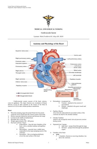
  • 1. Lecture Notes on Cardiovascular System Prepared By: Mark Fredderick R Abejo R.N, MAN MEDICAL AND SURGICAL NURSING Cardiovascular System Lecturer: Mark Fredderick R. Abejo RN, MAN Anatomy and Physiology of the Heart Cardiovascular system consists of the heart, arteries, veins & capillaries. The major function are circulation of blood, delivery of O2 & other nutrients to the tissues of the body & removal of CO2 & other cellular products metabolism Heart  Muscular pumping organ that propel blood into the arerial system & receive blood from the venous system of the body.  Hollow muscular behind the sternum and between the lungs  Located on the middle of mediastinum  Resemble like a close fist  Weighs approximately 300 – 400 grams  Has heart wall has 3 layers  Endocardium – lines the inner chambers of the heart, valves, chordate tendinae and papillary muscles.  Myocardium – muscular layer, middle layer, responsible for the major pumping action of the ventricles.  Epicardium – thin covering(mesothelium), covers the outer surface of the heart  Pericardium – invaginated sac  Visceral – attached to the exterior of myocardium  Parietal – attached to the great vessels and diaphragm  Papillary Muscle Arise from the endocardial & myocardial surface of the ventricles & attach to the chordae tendinae  Chordae Tendinae Attach to the tricuspid & mitral valves & prevent eversion during systole  Separated into 2 pumps:  right heart – pumps blood through the lungs  left heart – pumps blood through the peripheral organs  Chamber of the Heart Atria  2 chambers, function as receiving chambers, lies above the ventricles  Medical and Surgical Nursing 1 Abejo
  • 2. Lecture Notes on Cardiovascular System Prepared By: Mark Fredderick R Abejo R.N, MAN  Upper Chamber (connecting or receiving)  Right Atrium: receives systemic venous blood through the superior vena cava, inferior vena cava & coronary sinus  Left Atrium: receives oxygenated blood returning to the heart from the lungs trough the pulmonary veins Ventricles  2 thick-walled chambers; major responsibility for forcing blood out of the heart; lie below the atria  Lower Chamber (contracting or pumping)  Right Ventricle: contracts & propels deoxygenated blood into pulmonary circulation via the aorta during ventricular systole; Right atrium has decreased pressure which is 60 – 80 mmHg  Left Ventricle: propels blood into the systemic circulation via aortaduring ventricular systole; Left ventricle has increased pressure which is 120 – 180 mmHg in order to propel blood to the systemic circulation  Heart Valves  Tricuspid  Pulmonic  Mitral  Aortic  Coronary artery – 1st branch of aorta Right Coronary  SA nodal Branch – supplies SA node  Right marginal Branch – supplies the right border of the heart  AV nodal branch – supplies the AV node  Posterior interventricular artery – supplies both ventricles Left Coronary  Circumflex branch – supplies SA node in 40 % of people  Left marginal – supplies the left ventricle  Anterior interventricular branch aka Left anterior descending(LAD)–supplies both ventricles and interventricular septum  Lateral branch – terminates in ant surface of the heart  Coronary Veins Coronary sinus – main vein of the heart Great Cardiac vein – main tributary of the coronary sinus Oblique vein – remnant of SVC, small unsignificant Heart Circulation Cardiac Conduction System Properties of Heart Conduction System • Automaticity • Excitability • Conductivity • Contractility Structure of Heart Conduction System  Nodal tissues SA Node( Sino-atrial, Keith and Flack)  Primary Pacemaker  Between SVC and RA  Vagal and symphatetic innervation  Sinus Rhythms Medical and Surgical Nursing 2 Abejo
  • 3. Lecture Notes on Cardiovascular System Prepared By: Mark Fredderick R Abejo R.N, MAN AV Node( Atrioventricular , Kent and Tawara)  At the right atrium  3 zones  AN Zone(atrionodal)  N Zone (nodal)  NH zone (nodal –HIS)  Internodal and Interatrial Pathways Connects SA and AV Node Ant. Internodal(bachman) tract Middle Internodal(wenkebach) tract Posterior internodal(Thorel) tract  Bundle of His/ Purkinje Fibers Provides for ventricular conduction system Fastest conduction among cardiac tissues Right bundle Left Bundle Cardiac Action Potential  Depolarization: electrical activation of a cell caused by the influx of sodium into the cell while potassium exits the cell  Repolarization: return of the cell to the resting state caused by re-entry of potassium into the cell while sodium exits  Refractory periods: Effective refractory period: phase in which cells are incapable of depolarizing Relative refractory period: phase in which cells require a stronger-than-normal stimulus to depolarize Anatomical Sequence of Excitation of the Heart  (right atrium)  sinoatrial node (SA)  (right AV valve)  atrioventricular node (AV)  atrioventricular bundle (bundle of His)  right & left bundle of His branches  Purkinje fibers of ventricular walls (from SA through complete heart contraction = 220 ms = 0.22 s) a. Sinoatrial node (SA node) "the pacemaker" - has the fastest autorhythmic rate (70-80 per minute), and sets the pace for the entire heart; this rhythm is called the sinus rhythm; located in right atrial wall, just inferior to the superior vena cava b. Atrioventricular node (AV node) - impulses pass from SA via gap junctions in about 40 ms.; impulses are delayed about 100 ms to allow completion of the contraction of both atria; located just above tricuspid valve (between right atrium & ventricle) c. Atrioventricular bundle (bundle of His) - in the interATRIAL septum (connects L and R atria) d. L and R bundle of His branches - within the interVENTRICULAR septum (between L and R ventricles) e. Purkinje fibers - within the lateral walls of both the L and R ventricles; since left ventricle much larger, Purkinjes more elaborate here; Purkinje fibers innervate “papillary muscles” before ventricle walls so AV can valves prevent backflow The Normal Cardiac Cycle General Concepts Systole - period of chamber contraction Diastole - period of chamber relaxation Cardiac cycle - all events of systole and diastole during one heart flow cycle Events of Cardiac Cycle 1. mid-to-late ventricular diastole: ventricles filled  the AV valves are open  pressure: LOW in chambers; HIGH in aorta/pulmonary trunk  aortic/pulmonary semilunar valves CLOSED  blood flows from vena cavas/pulmonary vein INTO atria  blood flows through AV valves INTO ventricles (70%) 2. ventricular systole: blood ejected from heart  filled ventricles begin to contract, AV valves CLOSE  contraction of closed ventricles increases pressure  ventricular ejection phase - blood forced out  semilunar valves open, blood -> aorta & pulmonary trunk 3. isovolumetric relaxation: early ventricular diastole  ventricles relax, ventricular pressure becomes LOW  semilunar valves close, aorta & pulmonary trunk backflow TOTAL CARDIAC CYCLE TIME = 0.8 second (normal 70 beats/minute) atrial systole (contraction) = 0.1 second ventricular systole (contraction) = 0.3 second quiescent period (relaxation) = 0.4 second Cardiac Output - Blood Pumping of the Heart General Concepts • Stroke volume: the amount of blood ejected with each heartbeat • Cardiac output: amount of blood pumped by the ventricle in liters per minute • Preload: degree of stretch of the cardiac muscle fibers at the end of diastole • Contractility: ability of the cardiac muscle to shorten in response to an electrical impulse • Afterload: the resistance to ejection of blood from the ventricle • Ejection fraction: the percent of end-diastolic volume ejected with each heartbeat Medical and Surgical Nursing 3 Abejo
  • 4. Lecture Notes on Cardiovascular System Prepared By: Mark Fredderick R Abejo R.N, MAN General Variables of Cardiac Output 1. Cardiac Output (CO) - blood amount pumped per minute  CO (ml/min) = HR (beats/min) X SV (ml/beat)  Normal CO = 75 beats/min X 70 ml/beat = 5.25 L/min 2. Heart Rate (HR) - cardiac cycles per minute  Normal range is 60-100 beats per minute  Tachycardia is greater than 100 bpm  Bradycardia is less than 60 bpm  Sympathetic system INCREASES HR  Parasympathetic system (Vagus) DECREASES HR 3. Blood pressure - Cardiac output X peripheral resistance  Control is neural (central and peripheral) and hormonal  Baroreceptors in the carotid and aorta  Hormones- ADH, aldosterone, epinephrine can increase BP; ANF can decrease BP Regulation of Stroke Volume (SV)  End diastolic volume (EDV) - total blood collected in ventricle at end of diastole; determined by length of diastole and venous pressure (~ 120 ml)  End systolic volume (ESV) - blood left over in ventricle at end of contraction (not pumped out); determined by force of ventricle contraction and arterial blood pressure (~50 ml) SV (ml/beat) = EDV (ml/beat) - ESV (ml/beat) Normal SV = 120 ml/beat - 50 ml/beat = 70 ml/beat Frank-Starling Law of the Heart - critical factor for stroke volume is "degree of stretch of cardiac muscle cells"; more stretch = more contraction force increased EDV = more contraction force slow heart rate = more time to fill exercise = more venous blood return Regulation of Heart Rate (Autonomic, Chemical, Other) 1. Autonomic Regulation of Heart Rate (HR)  Sympathetic - NOREPINEPHRINE (NE) increases heart rate (maintains stroke volume which leads to increased Cardiac Output)  Parasympathetic - ACETYLCHOLINE (ACh) decreases heart rate  Vagal tone - parasympathetic inhibition of inherent rate of SA node, allowing normal HR  Baroreceptors, pressoreceptors - monitor changes in blood pressure and allow reflex activity with the autonomic nervous system 2. Hormonal and Chemical Regulation of Heart Rate (HR)  epinephrine - hormone released by adrenal medulla during stress; increases heart rate  thyroxine - hormone released by thyroid; increases heart rate in large quantities; amplifies effect of epinephrine  Ca++, K+, and Na+ levels very important; hyperkalemia - increased K+ level; KCl used to stop heart on lethal injection hypokalemia - lower K+ levels; leads to abnormal heart rate rhythms hypocalcemia - depresses heart function hypercalcemia - increases contraction phase hypernatremia - HIGH Na+ concentration; can block Na+ transport & muscle contraction 3. Other Factors Effecting Heart Rate (HR) normal heart rate - fetus 140 - 160 beats/minute female 72 - 80 beats/minute male 64 - 72 beats/minute 1. exercise - lowers resting heart rate (40-60) 2. heat - increases heart rate significantly 3. cold - decreases heart rate significantly 4. tachycardia - HIGHER than normal resting heart rate (over 100); may lead to fibrillation 5. bradycardia - LOWER than normal resting heart rate (below 60); parasympathetic drug side effects; physical conditioning; sign of pathology in non-healthy patient Vascular System Major function of the blood vessels isto supply the tissue with blood, remove wastes, & carry unoxygenated blood back to the heart Types of Blood Vessels Arteries Elastic-walled vessels that can stretch during systole & recoil during diastole; they carry blood away from the heart & distribute oxygenated blood throughout the body Arterioles Small arteries that distribute blood to the capillaries & function in controlling systemic vascular resistance & therefore arterial pressure Capilliaries The following exchanges occurs in the capilliaries O2 & CO2 Solutes between the blood & tissue Fluid volume transfer between the plasma & interstitial space Venules Small veins that receive blood from capillaries & function as collecting channels between the capillaries & veins Veins Low-pressure vessels with thin small & less muscles than arteries; most contains valves that prevent retrograde blood flow; they carry deoxygenated blood back to the heart. When the skeletal surrounding veins contract, the veins are compressed, promoting movement of blood back to the heart. Medical and Surgical Nursing 4 Abejo
  • 5. Lecture Notes on Cardiovascular System Prepared By: Mark Fredderick R Abejo R.N, MAN Assessment of the Client with Cardiovascular Disorders Nursing History Risk Factors A. Non – Modifiable Risk Factor  Age  Gender  Race  Heredity B. Modifiable Risk Factor  Stress  Diet  Exercise  Sedentary lifestyle  Cigarette smoking  Alcohol  Hypertension  Hyperlipidemia  DM  Obesity  Type A personality  Contraceptive Pills Common Clinical Manifestations of Cardiovascular Disorders a. Dyspnea - Exertional - Orthopnea - Paroxysmal Noctural Dyspnea - Cheyne-stokes b. Chest Pain c. Edema - Ascites - Hydrothorax - Anasarca d. Palpitation e. Hemoptysis f. Fatigue g. Syncope and Fainting h. Cyanosis i. Abdominal Pain j. Clubbing of fingers k. Jaundice Physical Assessment Inspection: – Skin color – Neck vein distention – Respirations – Pulsations – Clubbing – Capillary refill Palpation: Heart Sounds: Stethoscope Listening Overview of Heart Sounds (lub-du ; lub, dub ) lub - closure of AV valves, onset of ventricular systole dub - closure of semilunar valves, onset of diastole  Tricuspid valve (lub) - RT 5th intercostal, medial  Mitral valve (lub) - LT 5th intercostal, lateral  Aortic semilunar valve (dub) - RT 2nd intercostal  Pulmonary semilunar valve (dub) - LT 2nd intercostals S1 - due to closure of the AV(mitral/tricuspid) valves - timing: beginning of systole - loudest at the apex S2 - due to the closure of the semi-lunar (pulmonic/aortic) valves - timing: diastole - loudest at the base S3 – Ventricular Diastolic Gallop Mechanism: vibration resulting from resistance to rapid ventricular filling secondary to poor compliance Timing: early diastole Location: Apex (LV) or LLSB (RV) Pitch: faint and low pitched S4 - Atrial Diastolic Gallop Mechanism: vibration resulting from resistance to late ventricular filling during atrial systole Timing: late diastole ( before S1) Location: Apex ( LV) or LLSB (RV) Pitch: low ( use bell) Heart Murmurs Murmur - sounds other than the typical "lub-dub"; typically caused by disruptions in flow  Incompetent valve - swishing sound just AFTER the normal "lub" or "dub"; valve does not completely close, some regurgitation of blood  Stenotic valve - high pitched swishing sound when blood should be flowing through valve; narrowing of outlet in the open state Pericardial Friction Rub  It is an extra heart sound originating from the pericardial sac  Mechanism: Originates from the pericardial sac as it moves  Timing: with each heartbeat Medical and Surgical Nursing 5 Abejo
  • 6. Lecture Notes on Cardiovascular System Prepared By: Mark Fredderick R Abejo R.N, MAN  Location: over pericardium. Upright position, leaning forward  Pitch: high pitched and scratchy. Sounds like sandpaper being rubbed together  Significance: inflammation, infection, infiltration Classification of Clients with Diseases of the Heart ( Functional Capacity )  Class I. Patients with cardiac disease but without resulting limitations of physical activity.  Class II. Patients with cardiac disease resulting to slight limitation of physical activity  Class III. Patients with cardiac disease resulting in marked limitation of physical activity. They are comfortable at rest.  Class IV. Patients with cardiac disease resulting in inability to carry on any physical activity without discomfort Diagnostic Assessment Purposes: 1. To assist in diagnosing MI 2. To identify abnormalities 3. To assess inflammation 4. To determine baseline value 5. To monitor serum level of medications 6. To assess the effects of medications A. Blood Studies 1. Complete Blood Count a. RBC count- # of RBCs/ mm3 of blood, to diagnose anemia and ploycythemia b. Hemoglobin- # of grams of hgb/ 100ml of blood; to measure the oxygen-carrying capacity of the blood c. Hematocrit – expressed in %; measures the volume of RBCs in proportion to plasma; used also to diagnose anemia and polycythemia and abnormal hydration states d. RBC indices- measure RBC size and hemoglobin content a. MCV (mean corpuscular volume) b. MCH (mean corpuscular hemoglobin) c. MCHC (mean corpuscular hemoglobin concentrarion) e. Platelet count- # of Platelet/ mm3; to diagnose thrombocytopenia and subsequent bleeding tendencies f. WBC count- of WBCs/ mm3 of blood; to detect infection or inflammation g. WBC Differential count- determines proportion of each WBC in a sample of 100 WBCs; used to classify leukemias Normal Values RBC: Women – 4.2-5.4 million/mm3 Men – 4.7-6.1 million/mm3 Hgb: Women – 12-16 g/dl Men – 13-18 g/dl Hct : Women – 36-42% Men – 42-48% WBC: 5000-10,000/mm3 Granulocytes Neutrophils: 55-70% Eosinophils: 1-4% Basophils: 0.5-1.0% Agranulocytes Lymphocytes: 20-40% Monocytes: 2-8% Platelets: 150,000-450,000/mm3 2. Coagulation Screening Test a. Bleeding Time – measures the ability to stop bleeding after small puncture wound b. Partial Thromboplastin Time (PTT) – used to identify deficiencies of coagulation factors, prothrombin and fibrinogen; monitors heparin therapy. c. Prothrombin Time (Pro-time) – determines activity and interaction of the Prothrombin group: factors V (preacclerin), VII (proconvertin), X (Stuart-Power factor), prothrombin and fibrinogen; used to determine dosages of oral anti-coagulant. Normal Values Bleeding Time: 2.75-8 min Partial Thromboplastin Time (PTT): 60 - 70 sec. Prothrombin Time (PT): 12-14 sec. 3. Erythrocyte sedimentation rate ( ESR) It is a measurement of the rate at which RBC’s settle out of anticoagulated blood in an hour It is elevated in infectious heart disorder or myocardial infarction Normal Values Male: 15-20 mm/hr Female: 20-30 mm/hr 4. CARDIAC Proteins and enzymes a. CK- MB ( creatine kinase)  Most cardiac specific enzymes  Accurate indicator of myocardial dammage  Elevates in MI within 4 hours, peaks in 18 hours and then declines till 3 days  Normal value is 0-7 U/L or males 50-325 mu/ml Female 50-250 mu/ml b. Lactic Dehydrogenase (LDH)  Most sensitive indicator of myocardial damage  Elevates in MI in 24 hours, peaks in 48-72 hours Return to normal in 10-14 days  Normally LDH1 is greater than LDH2  Lactic Dehydrogenase (LDH)  MI- LDH2 greater than LDH1 (flipped LDH pattern)  Normal value is 70-200 IU/L (100 – 225 mu/ml) c. Myoglobin  Rises within 1-3 hours  Peaks in 4-12 hours  Returns to normal in a day  Not used alone  Muscular and RENAL disease can have elevated myoglobin d. Troponin I and T  Troponin I is usually utilized for MI  Elevates within 3-4 hours, peaks in 4-24 hours and persists for 7 days to 3 weeks!  Normal value for Troponin I is less than 0.6 ng/mL  REMEMBER to AVOID IM injections before obtaining blood sample!  Early and late diagnosis can be made! e. SERUM LIPIDS  Lipid profile measures the serum cholesterol, triglycerides and lipoprotein levels  Cholesterol= 200 mg/dL  Triglycerides- 40- 150 mg/dL  LDH- 130 mg/dL  HDL- 30-70- mg/dL  NPO post midnight (usually 12 hours) Medical and Surgical Nursing 6 Abejo
  • 7. Lecture Notes on Cardiovascular System Prepared By: Mark Fredderick R Abejo R.N, MAN B. Non-Invasive Procedure 1. Cardiac Monitoring / Electrocardiography (ECG) A non-invasive procedure that evaluates the electrical activity of the heart a. Limb Leads b. Precordial Leads The precordial leads VI –V6 are part of the 12 lead EKG. They are not monitored with the standard limb leads c. 12 lead ECG ECG Paper Deflection Waves of ECG 1. P wave - initial wave, demonstrates the depolarization from SA Node through both ATRIA; the ATRIA contract about 0.1 s after start of P Wave. 2. QRS complex - next series of deflections, demonstrates the depolarization of AV node through both ventricles; the ventricles contract throughout the period of the QRS complex, with a short delay after the end of atrial contraction; repolarization of atria also obscured 3. T Wave - repolarization of the ventricles (0.16 s) 4. PR (PQ) Interval - time period from beginning of atrial contraction to beginning of ventricular contraction (0.16 s) 5. QT Interval - the time of ventricular contraction (about 0.36 s); from beginning of ventricular depolarization to end of repolarization. 2. Holter Monitoring  A non-invasive test in which the client wears a Holter monitor and an ECG tracing recorded continuously over a period of 24 hours  Instruct the client to resume normal activities and maintain a diary of activities and any symptoms that may develop Medical and Surgical Nursing 7 Abejo
  • 8. Lecture Notes on Cardiovascular System Prepared By: Mark Fredderick R Abejo R.N, MAN 3. Stress Test  A non-invasive test that studies the heart during activity and detects and evaluates CAD  Exercise test, pharmacologic test and emotional test  Treadmill testing is the most commonly used stress test  Used to determine CAD, Chest pain causes, drug effects and dysrhythmias in exercise  Pre-test: consent may be required, adequate rest , eat a light meal or fast for 4 hours and avoid smoking, alcohol and caffeine  During the test: secure electrodes to appropriate location on chest, obtain baseline BP and ECG tracing, instruct client to exercise as instructed and report any pain, weakness and SOB, monitor BP and ECG continuously, record at frequent interval  Post-test: instruct client to notify the physician if any chest pain, dizziness or shortness of breath . Instruct client to avoid taking a hot shower for 10-12 hours after the test 4. Pharmacological stress test  Use of dipyridamole  Maximally dilates coronary artery  Side-effect: flushing of face  Pre-test: 4 hours fasting, avoid alcohol, caffeine  Post test: report symptoms of chest pain 5. ECHOCARDIOGRAM  Non-invasive test that studies the structural and functional changes of the heart with the use of ultrasound  Client Preparation: instruct client to remain still during the test, secure electrodes for simultaneous ECG tracing, explain that there will be no pain or electrical shock, lubricant placed on the skin will be cool. 6. Phonocardiography  Is a graphic recording of heart sound with simultaneous ECG. C. Invasive Procedure 1. Cardiac Catheterization ( Coronary Angiography / Arteriography )  Insertion of a catheter into the heart and surrounding vessels  Is an invasive procedure during which physician injects dye into coronary arteries and immediately takes a series of x-ray films to assess the structures of the arteries  Determines the structure and performance of the heart valves and surrounding vessels  Used to diagnose CAD, assess coronary atery patency and determine extent of atherosclerosis  Pretest: Ensure Consent, assess for allergy to seafood and iodine, NPO, document weight and height, baseline VS, blood tests and document the peripheral pulses  Pretest: Fasting for 8-12 hours, teachings, medications to allay anxiety  Intra-test: inform patient of a fluttery feeling as the catheter passes through the heart; inform the patient that a feeling of warmth and metallic taste may occur when dye is administered  Post-test: Monitor VS and cardiac rhythm  Monitor peripheral pulses, color and warmth and sensation of the extremity distal to insertion site  Maintain sandbag to the insertion site if required to maintain pressure  Monitor for bleeding and hematoma formation 2. Nuclear Cardiology  Are safe methods of evaluating left ventricular muscle function and coronary artery blood distribution.  Client Preparation: obtain written consent, explain procedure, instruct client that fasting may be required for a short period before the exam, assess for iodine allergy.  Post Procedure: encourage client to drink fluids to facilitate the excretion of contrast material, assess venipuncture site for bleeding or hematoma.  Types of Nuclear Cardiology o Multigated acquisition (MUGA) or cardiac blood pool scan  Provides information on wall motion during systole and diastole, cardiac valves, and EF. o Single-photon emission computed tomography (SPECT)  Used to evaluate the myocardium at risk of infarction and to determine infarction size. o Positron emission tomography (PET) scanning  Uses two isotopes to distinguish viable and nonviable myocardial tissue. Medical and Surgical Nursing 8 Abejo
  • 9. Lecture Notes on Cardiovascular System Prepared By: Mark Fredderick R Abejo R.N, MAN o Perfusion imaging with exercise testing  Determines whether the coronary blood flow changes with increased activity.  Used to diagnose CAD, determine the prognosis in already diagnosed CAD, assess the physiologic significance of a known coronary lesion, and assess the effectiveness of various therapeutic modalities such as coronary artery bypass surgery, percutaneous coronary intervention, or thrombolytic therapy. D. Hemodynamics Monitoring 1. CVP ( Central Venous Pressure )  Reflects the pressure of the blood in the right atrium.  Engorgement is estimated by the venous column that can be observed as it rises from an imagined angle at th point of manubrium ( angle of Louis).  With normal physiologic condition, the jugular venous column rises no higher than 2-3 cm above the clavicle with the client in a sitting position at 45 degree angle.  CVP is a measurement of: - cardiac efficiency - blood volume - peripheral resistance  Right ventricular pressure – a catheter is passed from a cutdown in the antecubital, subclavian jugular or basilica vein to the right atrium and attached to a prescribed manometer or tranducer.  NORMAL CVP is 2 -8 cm h20 or 2-6 mm Hg  Decrease indicates dec. circulating volume, increase indicates inc. blood volume or right heart beat failure.  To Measure: patient should be flat with zero point of manometer at the same level of the RA which corresponds to the mid-axillary line of the patient or approx. 5 cm below the sternum.  Fluctuations follow patients respiratory function and will fall on inspiration and rise on expiration due to changes in intrapulmonary pressure. Reading should be obtained at the highest point of fluctuation. 2. Pulmonary Artery Pressure ( PAP) Monitoring  Appropriate for critically ill clients requiring more accurate assessments of the left heart pressure  Swan-Ganz Catheter / Pulmonary Artery Catheter is use  Client Preparation: obtain consent, insertion is under strict sterile technique, usually at the bedside, explain to client the sterile drapes may cover the face, assists to position client flat or slight T-postion as tolerated and instruct to remain still during the procedure  Nursing Care During Insertion: Monitor and document HR,BP and ECG during the procedure CARDIAC DISORDER CORONARY ARTERIAL DISEASE ISCHEMIC HEART DISEASE Results from the focal narrowing of the large and medium-sized coronary arteries due to deposition of atheromatous plaque in the vessel wall Stages of Development of Coronary Artery Disease 1. Myocardial Injury: Atherosclerosis 2. Myocardial Ischemia: Angina Pectoris 3. Myocardial Necrosis: Myocardial Infarction I. ATHEROSCLEROSIS ATHEROSCLEROSIS ARTERIOSCLEROSIS Narrowing of artery Lipid or fat deposits Tunica intima Hardening of artery Calcium and protein deposits Tunica media A. PRESDISPOSING FACTORS 1. Sex: male 2. Race: black 3. Smoking 4. Obesity 5. Hyperlipidemia 6. Sedentary lifestyle 7. Diabetes Mellitus 8. Hypothyroidism 9. Diet: increased saturated fats 10. Type A personality B. SIGNS AND SYMPTOMS 1. Chest pain 2. Dyspnea 3. Tachycardia 4. Palpitations 5. Diaphoresis C. TREATMENT Percutaneous Transluminal Coronary Angioplasty and Intravascular Stenting  Mechanical dilation of the coronary vessel wall by compresing the atheromatous plaque.  It is recommended for clients with single-vessel coronary artery disease. Medical and Surgical Nursing 9 Abejo
  • 10. Lecture Notes on Cardiovascular System Prepared By: Mark Fredderick R Abejo R.N, MAN  Prosthetic intravascular cylindric stent maintain good luminal geometry after ballon deflation and withdrawal.  Intravascular stenting is done to prevent restenosis after PTCA Coronary Arterial Bypass Graft Surgery Greater and lesser saphenous veins are commonly used for bypass graft procedures Objectives of CABG 1. Revascularize myocardium 2. To prevent angina 3. Increase survival rate 4. Done to single occluded vessels 5. If there is 2 or more occluded blood vessels CABG is done Nursing Management:  Nitroglycerine is the drug of choice for relief of pain from acute ischemic attacks  Instruct to avoid over fatigue  Plan regular activity program For Saphenous Vein Site:  Wear support stocking 4-6 week postop  Apply pressure dressing or sand bag on the site  Keep leg elevated when sitting 3 Complications of CABG 1. Pneumonia: encourage to perform deep breathing, coughing exercise and use of incentive spirometer 2. Shock 3. Thrombophlebitis II. ANGINA PECTORIS Transient paroxysmal chest pain produced by insufficient blood flow to the myocardium resulting to myocardial ischemia Clinical syndrome characterized by paroxysmal chest pain that is usually relieved by rest or nitroglycerine due to temporary myocardial ischemia Types of Angina Pectoris  Stable Angina: pain less than 15 minutes, recurrence is less frequent.  Unstable Angina : pain is more than 15 mins.,but not less than 30 minutes, recurrence is more frequent and the intensity of pain increases.  Variant Angina ( Prinzmetal’s Angina ): Chest pain is on longer duration and may occur at rest. Result from coronary vasospasm.  Angina Decubitus: paroxysmal chest pain that occur when the client sits or stand. A. PRESDISPOSING FACTORS 1. Sex: male 2. Race: black 3. Smoking 4. Obesity 5. Hyperlipidemia 6. Sedentary lifestyle 7. Diabetes Mellitus 8. Hypertension 9. CAD: Atherosclerosis 10. Thromboangiitis Obliterans 11. Severe Anemia 12. Aortic Insufficiency: heart valve that fails to open & close efficiently 13. Hypothyroidism 14. Diet: increased saturated fats 15. Type A personality B. PRESIPITATING FACTORS 4 E’s of Angina Pectoris 1. Excessive physical exertion: heavy exercises, sexual activity 2. Exposure to cold environment: vasoconstriction 3. Extreme emotional response: fear, anxiety, excitement, strong emotions 4. Excessive intake of foods or heavy meal C. SIGNS AND SYMPTOMS 1. Levine’s Sign: initial sign that shows the hand clutching the chest 2. Chest pain: characterized by sharp stabbing pain located at sub sterna usually radiates from neck, back, arms, shoulder and jaw muscles usually relieved by rest or taking nitroglycerine(NTG) 3. Dyspnea 4. Tachycardia 5. Palpitations 6. Diaphoresis Medical and Surgical Nursing 10 Abejo
  • 11. Lecture Notes on Cardiovascular System Prepared By: Mark Fredderick R Abejo R.N, MAN D. DIAGNOSTIC PROCEDURE 1. History taking and physical exam 2. ECG: may reveals ST segment depression & T wave inversion during chest pain 3. Stress test / treadmill test: reveal abnormal ECG during exercise 4. Increase serum lipid levels 5. Serum cholesterol & uric acid is increased E. MEDICAL MANAGEMENT 1. Drug Therapy: if cholesterol is elevated Nitrates: Nitroglycerine (NTG) Beta-adrenergic blocking agent: Propanolol Calcium-blocking agent: nefedipine Ace Inhibitor: Enapril 2. Modification of diet & other risk factors 3. Surgery: Coronary artery bypass surgery 4. Percutaneuos Transluminal Coronary Angioplasty (PTCA) F. NURSING INTERVENTIONS 1. Enforce complete bed rest 2. Give prompt pain relievers with nitrates or narcotic analgesic as ordered 3. Administer medications as ordered: A. Nitroglycerine(NTG): when given in small doses will act as venodilator, but in large doses will act as vasodilator  Give 1st dose of NTG: sublingual 3-5 minutes  Give 2nd dose of NTG: if pain persist after giving 1st dose with interval of 3-5 minutes  Give 3rd& last dose of NTG: if pain still persist at 3-5 minutes interval NTG Tablets(sublingual)  Keep the drug in a dry place, avoid moisture and exposure to sunlight as it may inactivate the drug  Change stock every 6 months  Offer sips of water before giving sublingual nitrates, dryness of mouth may inhibit drug absoprtion  Relax for 15 minutes after taking a tablet: to prevent dizziness  Monitor side effects: orthostatic hypotension, flushed face. Transient headache & dizziness: frequent side effect  Instruct the client to rise slowly from sitting position  Assist or supervise in ambulation NTG Nitrol or Transdermal patch  Nitropatch is applied once a day, usually in the morning.  Avoid placing near hairy areas as it may decrease drug absorption  Avoid rotating transdermal patches as it may decrease drug absorption  Avoid placing near microwave ovens or during defibrillation as it may lead to burns (most important thing to remember) B. Beta-blockers: decreases myocardial oxygen demand by decreasing heart rate, cardiac output and BP Propanolol Metropolol Pindolol Atenolol  Assess PR, withhold if dec.PR  Administer with food ( prevent GI upset )  Propanolol: not given to COPD cases: it causes bronchospasm and DM cases: it cause hypoglycemia  Side Effects: Nausea and vomiting, mental depression and fatigue C. Calcium – Channel Blockers: relaxes smooth cardiac muscle, reduces coronary vasospasm Amlodipine ( norvasc ) Nifedipine ( calcibloc ) Diltiazem ( cardizem )  Assess HR and BP  Adminester 1 hour before meal and 2 hours after meal ( foods delay absorption ) 4. Administer oxygen inhalation 5. Place client on semi-to high fowlers position 6. Monitor strictly V/S, I&O, status of cardiopulmonary fuction & ECG tracing 7. Provide decrease saturated fats sodium and caffeine 8. Provide client health teachings and discharge planning  Avoidance of 4 E’s  Prevent complication (myocardial infarction)  Instruct client to take medication before indulging into physical exertion to achieve the maximum therapeutic effect of drug  Reduce stress & anxiety: relaxation techniques & guided imagery  Avoid overexertion & smoking  Avoid extremes of temperature  Dress warmly in cold weather  Participate in regular exercise program  Space exercise periods & allow for rest periods  The importance of follow up care 9. Instruct the client to notify the physician immediately if pain occurs & persists despite rest & medication administration III. MYOCARDIAL INFARCTION Death of myocardial cells from inadequate oxygenation, often caused by sudden complete blockage of a coronary artery Characterized by localized formation of necrosis (tissue destruction) with subsequent healing by scar formation & fibrosis Heart attack Terminal stage of coronary artery disease characterized by malocclusion, necrosis & scarring. Types of M.I  Transmural Myocardial Infarction: most dangerous type characterized by occlusion of both right and left coronary artery  Subendocardial Myocardial Infarction: characterized by occlusion of either right or left coronary artery The Most Critical Period Following Diagnosis of Myocardial Infarction 6-8 hours because majority of death occurs due to arrhythmia leading to premature ventricular contractions (PVC) A. PREDISPOSING FACTORS 1. Sex: male 2. Race: black 3. Smoking 4. Obesity 5. CAD: Atherosclerotic 6. Thrombus Formation 7. Genetic Predisposition 8. Hyperlipidemia Medical and Surgical Nursing 11 Abejo
  • 12. Lecture Notes on Cardiovascular System Prepared By: Mark Fredderick R Abejo R.N, MAN 9. Sedentary lifestyle 10. Diabetes Mellitus 11. Hypothyroidism 12. Diet: increased saturated fats 13. Type A personality B. SIGNS AND SYMPTOMS 1. Chest pain Excruciating visceral, viselike pain with sudden onset located at substernal& rarely in precordial Usually radiates from neck, back, shoulder, arms, jaw & abdominal muscles (abdominal ischemia): severe crushing Not usually relieved by rest or by nitroglycerine 2. N/V 3. Dyspnea 4. Increase in blood pressure & pulse, with gradual drop in blood pressure (initial sign) 5. Hyperthermia: elevated temp 6. Skin: cool, clammy, ashen 7. Mild restlessness & apprehension 8. Occasional findings: Pericardial friction rub Split S1& S2 Rales or Crackles upon auscultation S4 or atrial gallop C. DIAGNOSTIC PROCEDURED 1. Cardiac Enzymes CPK-MB: elevated Creatinine phosphokinase(CPK):elevated Heart only, 12 – 24 hours Lactic acid dehydrogenase(LDH): is increased Serum glutamic pyruvate transaminase(SGPT): is increased Serum glutamic oxal-acetic transaminase(SGOT): is increased 2. Troponin Test: is increased 3. ECG tracing reveals ST segment elevation T wave inversion Widening of QRS complexes: indicates that there is arrhythmia in MI 4. Serum Cholesterol & uric acid: are both increased 5. CBC: increased WBC D. NURSING INTERVENTIONS Goal: Decrease myocardial oxygen demand 1. Decrease myocardial workload (rest heart) Establish a patent IV line Administer narcotic analgesic as ordered: Morphine Sulfate IV: provide pain relief(given IV because after an infarction there is poor peripheral perfusion & because serum enzyme would be affected by IM injection as ordered) Side Effects: Respiratory Depression Antidote: Naloxone (Narcan) Side Effects of Naloxone Toxicity: is tremors 2. Administer oxygen low flow 2-3 L / min: to prevent respiratory arrest or dyspnea & prevent arrhythmias 3. Enforce CBR in semi-fowlers position without bathroom privileges(use bedside commode): to decrease cardiac workload 4. Instruct client to avoid forms of valsalva maneuver 5. Place client on semi fowlers position 6. Monitor strictly V/S, I&O, ECG tracing & hemodynamic procedures 7. Perform complete lung / cardiovascular assessment 8. Monitor urinary output & report output of less than 30 ml / hr: indicates decrease cardiac output 9. Provide a full liquid diet with gradual increase to soft diet: low in saturated fats, Na & caffeine 10. Maintain quiet environment 11. Administer stool softeners as ordered:to facilitate bowel evacuation & prevent straining 12. Relieve anxiety associated with coronary care unit(CCU)environment 13. Administer medication as ordered: a. Vasodilators:Nitroglycirine (NTG), Isosorbide Dinitrate, Isodil (ISD): sublingual b. Anti Arrythmic Agents: Lidocaine (Xylocane), Brithylium Side Effects: confusion and dizziness c. Beta-blockers: Propanolol (Inderal) d. ACE Inhibitors: Captopril (Enalapril) e. Calcium Antagonist: Nefedipine f. Thrombolytics / Fibrinolytic Agents: Streptokinase, Urokinase, Tissue Plasminogen Activating Factor (TIPAF) Side Effects:allergic reaction, urticaria, pruritus Nursing Intervention: Monitor for bleeding time g. Anti Coagulant Heparin Antidote: Protamine Sulfate Nursing Intervention: Check for Partial Thrombin Time (PTT) Caumadin(Warfarin) Antidote:Vitamin K Nursing Intervention: Check for Prothrombin Time (PT) h. Anti Platelet: PASA (Aspirin): Anti thrombotic effect Side Effects:Tinnitus, Heartburn, Indigestion / Dyspepsia Contraindication:Dengue, Peptic Ulcer Disease, Unknown cause of headache 14. Provide client health teaching & discharge planning concerning: a. Effects of MI healing process & treatment regimen b. Medication regimen including time name purpose, schedule, dosage, side effects c. Dietary restrictions: low Na, low cholesterol, avoidance of caffeine d. Encourage client to take 20 – 30 cc/week of wine, whisky and brandy:to induce vasodilation e. Avoidance of modifiable risk factors f. Prevent Complication Arrhythmia: caused by premature ventricular contraction Cardiogenic shock: late sign is oliguria Left Congestive Heart Failure Thrombophlebitis: homan’s sign Stroke / CVA Dressler’s Syndrome(Post MI Syndrome):client is resistant to pharmacological agents: administer 150,000-450,000 units of streptokinase as ordered g. Importance of participation in a progressive activity program h. Resumption of ADL particularly sexual intercourse: is 4-6 weeks post cardiac rehab, post CABG & instruct to: Medical and Surgical Nursing 12 Abejo
  • 13. Lecture Notes on Cardiovascular System Prepared By: Mark Fredderick R Abejo R.N, MAN Make sex as an appetizer rather than dessert Instruct client to assume a non weight bearing position Client can resume sexual intercourse: if can climb or use the staircase i. Need to report the ff s/sx: Increased persistent chest pain Dyspnea Weakness Fatigue Persistent palpitation Light headedness j. Enrollment of client in a cardiac rehabilitation program k. Strict compliance to mediation & importance of follow up care IV. CARDIOGENIC SHOCK ( POWER/PUMP FAILURE ) Is a shock state which result from profound left ventricular failure usually from massive MI. It result to low cardiac output, thereby systemic hypoperfusion. A. SIGNS AND SYMPTOMS 1. Decrease systolic BP 2. Oliguria 3. Cold, clammy skin 4. Weak pulse 5. Cyanosis 6. Mental lethargy 7. Confusion B. MEDICAL MANAGEMENT 1. Counterpulsation ( mechanical cardiac assistance / diastolic augmentation ) Involves introduction of the intra – aortic balloon catheter via the femoral artery Intra Aortic Balloon Pump augments diastole, resulting in increased perfusion of the coronary arteries and the myocardium and a decrease in left ventricular workload. The balloon is inflated during diastole, it is deflated during sytole. Indications:  Cardiogenic shock  AMI  Unstable Angina  Open heart surgery C. NURSING INTERVENTIONS 1. Perform hemodynamic monitoring 2. Administer oxygen therapy 3. Correct hypovolemia. Administer IV fluids as ordered 4. Pharmacology: a. Vasodilators: Nitroglycerine b. Inotropic agents:Digitalis, Dopamine c. Diuretics : Furosemide d. Sodium Bicarbonate, Relieve lactic acidosis 5. Monitor hourly urine output, LOC and arrhythmias 6. Provide psychosocial support 7. Decrease pulmonary edema a. Auscultate lung fields for crackles and wheezes b. Note for dyspnea, cough , hemoptysis and orthopnea c. Monitor ABG for hypoxia and metabolic acidosis d. Place in fowler’s position to reduce venous return e. Administer during therapy as ordered: Morphine sulfate to reduce venous return. Aminophylline to reduce bronchospasm caused by severe congestion. Vasodilators to reduce venous return Diuretics to decrease circulating volume V. PERICARDITIS / DRESSLER’S SYNDROME Is the inflammation of the pericardium which occurs approximately 1 – 6 weeks after AMI. Results as an antigen – antibody response. The necrotic tissues play the role of an antigen, which trigger antibody formation. Inflammatory process follows. Constrictive Pericarditis is a condition in which a chronic inflammatory thickening of the pericardium compresses the heart so that it is unable to fill normally during diastole. A. SIGNS AND SYMPTOMS 1. Pain in the anterior chest, aggravated by coughing, yawning, swallowing, twisting and turning the torso, relieved by upright, leaning forward position. 2. Pericardial friction rub – scratchy, grating or cracking sound 3. Dyspnea 4. Fever, sweating, chills 5. Joints pains 6. Arrhythmias B. NURSING INTERVENTIONS 1. Elevate head of bed, place pillow on the overbed table so that the patient can lean on it. 2. Bed rest 3. Administer prescribed pharmacotherapy. a. ASA to suppress inflammatory process b. Corticosteriods for more severe symptoms 4. Assist in pericardiocentesis if cardiac tamponade is present. 5. Pericardiocentesis is aspiration of blood or fluid from pericardial sac. VI. CARDIAC TAMPONADE Also known as pericardial tamponade, is an emergency condition in which fluid accumulates in the pericardium (the sac in which the heart is enclosed). If the fluid significantly elevates the pressure on the heart it will prevent the heart's ventricles from filling properly. This in turn leads to a low stroke volume. The end result is ineffective pumping of blood, shock, and often death. A. PREDISPOSING FACTORS 1. Chest trauma ( blunt or penetrating ) 2. Myocardial ruptured 3. Cancer 4. Pericarditis 5. Cardiac surgery ( first 24 – 48 hours ) 6. Thrombolytic therapy B. SIGNS AND SYMPTOMS 1. Beck’s Triad  Hypotension  Jugular venous distension  Muffled heart sound 2. Pulsus paradoxus ( drop of at least 10 mmHg in arterial BP on inspiration ) 3. Tachycardia 4. Breathlessness 5. Decrease in LOC Medical and Surgical Nursing 13 Abejo
  • 14. Lecture Notes on Cardiovascular System Prepared By: Mark Fredderick R Abejo R.N, MAN C. NURSING INTERVENTIONS 1. Administer oxygen 2. Elevate head of bed, place pillow on the overbed table so that the patient can lean on it. 3. Bed rest 4. Administer prescribed pharmacotherapy. c. ASA to suppress inflammatory process d. Corticosteriods for more severe symptoms 5. Assist in pericardiocentesis and thoracotomy 6. Pericardiocentesis is aspiration of blood or fluid from pericardial sac. CONGESTIVE HEART FAILURE Inability of the heart to pump blood towards systemic circulation I. LEFT-SIDED HEART FAILURE A. PREDISPOSING FACTORS 1. 90% - Mitral valve stenosis  RHD  Inflammation of mitral valve  Anti-streptolysin O titer (ASO) – 300 todd units  Penicillin, PASA, steroids  Aging 2. MI 3. IHD 4. HPN 5. Aortic valve stenosis B. SIGNS AND SYMPTOMS 1. Pulmonary edema/congestion  Dyspnea, PND (awakening at night d/t difficulty in breathing), 2-3 pillow orthopnea  Productive cough (blood tinged)  Rales/crackles  Bronchial wheezing  Frothy salivation 2. Pulsus alternans (A unique pattern during which the amplitude of the pulse changes or alternates in size with a stable heart rhythm.)This is common in severe left ventricular dysfunction.) 3. Anorexia and general body malaise 4. PMI displaced laterally, cardiomegaly 5. S3 (ventricular gallop) C. DIAGNOSTICS 1. CXR – cardiomegaly 2. PAP – pulmonary arterial pressure  Measures pressure in right ventricle  Reveals cardiac status 3. PCWP – pulmonary capillary wedge pressure  Measures end-systolic and end-diastolic pressure (elevated)  Done through cardiac catheterization (Swan- Ganz) 4. Echocardiograph – reveals enlarged heart chamber 5. ABG analysis reveals elevated PCO2 and decreased PO2 (respiratory acidosis)  hypoxemia and cyanosis Tracheostomy  for severe respiratory distress and laryngospasm  performed at bedside within 10-15 minutes CVP  reveals fluid status; Normal = 4-10cm H2o; right atrium PAP – cardiac status; left atrium ALLEN’S test – collateral circulation Cardiac Tamponade: pulsus paradoxus, muffled heart sounds, HPN II. RIGHT SIDED HEART FAILURE A. PREDISPOSING FACTORS 1. Tricuspid valve stenosis 2. COPD 3. Pulmonary embolism (char by chest pain and dyspnea) 4. Pulmonic stenosis 5. Left sided heart failure B. SIGNS AND SYMPTOMS (Venous congestion) 1. Jugular vein distention 2. Pitting edema 3. Ascites 4. Weight gain 5. Hepatosplenomegaly 6. Jaundice 7. Pruritus/ urticaria 8. Esophageal varices 9. Anorexia 10. Generalized body malaise C. DIAGNOSTICS 1. CXR – cardiomegaly 2. CVP – measures pressure in right atrium; N = 4- 10cc H2O  During CVP: trendelenburg  to prevent pulmo embolism and to promote ventricular filling  Flat on bed post CVP, check CVP readings  Hypovolemia – fluid challenge  Hypervolemia – diuretics (loop) 3. Echocardiography – reveals enlarged heart chamber  Muffled heart sounds  cardiomyopathy  Cyanotic heart diseases  TOF  “tet” spells  cyanosis with hypoxemia  Tricuspid valve stenosis  Transposition of aorta  Acyanotic  PDA – machine-like murmur  DOC: indomethacin SE: corneal cloudiness 4. Liver enzymes  SGPT up  SGOT up D. NURSING MANAGEMENT Goal: increase myocardial contraction  increase CO; Normal CO is 3-6L/min; N stroke volume is 60-70ml/h2o 1. Administer medications as ordered  Cardiac glycosides  Digoxin (N=.5-1.5, tox=2)  Tox: Anorexia, N&V; A: Digibind  Digitoxin – given if (+) ARF; metabolized in liver and not in kidneys  Loop diuretics  Lasix – IV push, mornings  Bronchodilators  Aminophylline (theophylline)  Tachycardia, palpitations  CNS hyperactivity, agitation  Narcotic analgesics  Morphine sulfate – induces vasodilation  Vasodilators  NTG and ISDN  Anti-arrhythmic agents  Lidocaine (SE: dizziness and confusion)  Bretyllium  YOU DON’T GIVE BETA-BLOCKERS TO THESE PATIENTS 2. Administer O2 inhalation at 3-4 L/minute via NC as ordered  high flow 3. High fowler’s, 2-3 Pillows 4. Restrict Na and fluids 5. Monitor strictly VS and IO and Breath Sounds 6. Weigh pt daily and assess for pitting edema 7. abdominal girth daily and notify MD 8. provide meticulous skin care Medical and Surgical Nursing 14 Abejo
  • 15. Lecture Notes on Cardiovascular System Prepared By: Mark Fredderick R Abejo R.N, MAN 9. provide a dietary intake which is low in saturated fats and caffeine 10. Institute bloodless phlebotomy  ROTATING TOURNIQUET  Rotated clockwise every 15 minutes to promote a decrease in venous return 11. Health teaching and discharge planning  Prevent complications : Arrhythmia, Shock, Thrombophlebitis, MI, Cor pulmonale – RV hypertrophy  Regular adherence to medications  Diet modifications  Importance of ffup care HYPERTENSION Is an abnormal elevation of Bp, systolic pressure above 140 mmHg and or diastolic pressure above 90mmHg at least two readings WHO: BP >160/95 mmHg AHA: BP >140/90 mmHg In hypertension, vasoconstriction – vasospasm – increases PVR – decrease blood flow to the organ. Target Organs:  Heart : MI, CHF, Dysrhythmias  Eyes: blurred / impaired vision, retinopathy, cataract.  Brain: CVA, encephalopathy  Kidneys : renal insufficiency, RF  Peripheral Bloods Vessels – aneurysm, gangrene CLASSIFICATION OF BP FOR ADULTS 18 YRS AND OLDER (PHIL. SOCIETY OF HPN) Optimal o <120 mmHg / <80 mmHg Recheck in 2 years. Normal o 120-129 mmHg / 80-84 mmHg Recheck in 2 years. High normal o 130-139 mmHg / 85-89 mmHg Recheck in 1 year. Stage 1 (mild) HPN o 140-159 mmHg / 90-99 mmHg Confirm in 2 months. Stage 2 (moderate) HPN o 160-179 mmHg / 100-109 mmHg Evaluate within a month. Stage 3 (severe) HPN o 180-209 mmHg / 110-119mmHg Evaluate within a week. Stage 4 (very severe) HPN o 210 mmHg / >/=120 mmHg Evaluate A. CLASSIFICATION  Essential / Idiophatic / Primary HPN, accounts for 90 – 95% of all cases of HPN, cause is unknown  Secondary HPN, due to known causes ( Renal failure, Hypertension )  Malignant Hypertension, is severe, rapidly progressive elevation in BP that causes rapid onset of end organ complication  Labile HPN, intermittently elevated BP  Resistant HPN, does not respond to usual treatment  White Coat HPN, elevation of B only during clinic or hospital visits  Hypertensive Crisis, situation that requires immediate blood pressure lowering 240mmHg / 120 mmHg B. RISK FACTORS 1. Family history 2. Age 3. High salt intake 4. Low potassium intake 5. Obesity 6. Excess alcohol consumption 7. Smoking 8. Stress C. SIGNS AND SYMPTOMS 1. Headache 2. Epistaxis 3. Dizziness 4. Tinnitus 5. Unsteadiness 6. Blurred vision 7. Depression 8. Nocturia 9. Retinopathy D. TREATMENT STRATEGIES Non-pharmacologic therapy 1. Low salt diet. 2. Weight reduction. 3. Exercise. 4. Cessation of smoking. 5. Decreased alcohol consumption. 6. Psychological methods: Relaxation / meditation. 7. Dietary decrease in saturated fat. Drug therapy Stepped Care o Progressive addition of drugs to a regimen, starting with one, usually a diuretic, and adding, in a stepwise fashion, a sympatholytic, vasodilator, and sometimes an ACE inhibitor. Monotherapy o Advantageous because of its simplicity, better patient compliance, and relatively low incidence of toxicity. CATEGORIES OF ANTI-HYPERTENSIVE DRUGS Drugs that alter sodium and water balance  Diuretics. Loop diuretics Thiazides Spironolactone and Triamterene Drugs that alter sympathetic nervous system function  Sympatholytic drugs. Centrally-acting sympatholytics  Clonidine  Guanabenz  Guanfacine  Methyldopa Peripherally-acting sympatholytics  Guanadrel  Guanethidine  Reserpine a-blockers  Doxazosin  Prazosin b-blockers  Acebutolol - Labetalol  Atenolol - Metoprolol  Betaxolol - Nadolol  Bisoprolol - Penbutolol  Carteolol - Pindolol  Carvedilol - Propranolol  Esmolol - Timolol Medical and Surgical Nursing 15 Abejo
  • 16. Lecture Notes on Cardiovascular System Prepared By: Mark Fredderick R Abejo R.N, MAN Vasodilators Direct vasodilators  Diazoxide - Hydralazine  Minoxidil - Nitroprusside  Fenoldopam Calcium channel blockers  Amlodipine - Nifedipine  Diltiazem - Nimodipine  Felodipine - Nisoldipine  Isradipine - Nitrendipine  Manidipine - Nicardipine  Lacidipine - Verapamil  Lercanidipine - Gallopamil AGENTS THAT BLOCK THE PRODUCTION OR ACTION OF ANGIOTENSIN  ACE inhibitors  Benazepril - Moexipril  Captopril - Quinapril  Enalapril - Perindopril  Fosinopril - Ramipril  Lisinopril - Trandolapril AT1-receptor blockers  Irbesartan - Losartan  Telmisartan - Valsartan  Candesartan - Eprosartan  Olmesartan DRUGS FOR HYPERTENSIVE EMERGENCIES OR CRISES Trimethaphan o 1 mg/ml IV infusion; titrate; instantaneous onset Sodium nitroprusside o 5-10 mg/L IV infusion; titrate; instantaneous onset Diazoxide o 300-600 mg Rapid IV push; instantaneous onset Nifedipine o 10-20 mg Sublingual or chewed; onset within 5-30 min. Labetalol o 20-80 mg IV at 10-minute intervals (max.dose: 300mg); immediate onset MECHANISMS OF DRUG ACTION PRINCIPLES OF DRUG THERAPY Monotherapy is generally reserved for mild to moderate HPN; it has gained popularity because of its simplicity, fewer side effects, and improved patient compliance. More severe HPN may require treatment with several drugs that are selected to minimize adverse effects of combined regimen. Treatment is initiated with any of 4 drugs depending on individual patient: Diuretic, b-blocker, ACEI, and a Ca-channel blocker; if BP is inadequately controlled, a 2nd-drug is then added. HPN may co-exist with other disease that may be aggravated by some of the anti-HPN agents. Lack of patient compliance is the most common reason for failure of anti-HPN therapy; it is important to enhance compliance by carefully selecting a drug regimen that minimizes adverse effects. Therapy is directed at preventing disease that may occur in the future, rather than in relieving present discomfort of the patient. Medical and Surgical Nursing 16 Abejo
  • 17. Lecture Notes on Cardiovascular System Prepared By: Mark Fredderick R Abejo R.N, MAN E. NURSING INTERVNTIONS 1. Patient Teaching and Counselling  Teaching about HPN and its risk factors  Stress therapy  Low NA and low saturated fat  Avoid stimulants ( caffeine, alcohol, smoking )  Regular pattern of exercise  Weight reduction if obese 2. Teaching about medication  The most common side effects of diuretics are potassium depletion and orthostatic hypotension.  The most common side effect of the different antihypertensive drugs is orthostatic hypotension.  Take anti – hypertensive medications at regular basis  Assume sitting or lying position for few minutes  Avoid very warm bath  Avoid prolonged sitting and standing  Avoid alcoholic beverages  Avoid tyramine – rich foods ( proteins ) as follows: ( this may cause hypertensive crisis ) Aged cheese Liver Beer Wine Chocolate Pickles Sausages Soy sauce 3. Preventing Non-compliance  Inform the client that absence of symptoms does not indicate control of BP  Advise the client against abrupt withdrawal of medication, rebound hypertension may occur.  Device ways to facilitate remembering of taking medications PERIPHERAL VASCULAR DISORDERS ANEURYSM It is the localized, irreversible dilatation of an artery secondary to an alteration in the integrity of its wall. Most common type is AAA ( abdominal aortic aneurysm ) The most common cause is hypertension A. CLASSIFICATIONS  Fusiform Aneurysm , involves outpouching of the both side of the artery  Saccular Aneurysm , outpouching of only one side of the artery.  Dissecting Aneurysm, involves separation or tear in the tunica intima and tunica media B. RISK FACTOR 1. Age 2. Tobacco use 3. HPN 4. Atherosclerosis 5. Race 6. Gender 7. Family history C. SIGNS AND SYMPTOMS 1. Pulsating mass over abdomen (AAA) 2. Presence of the bruit sound 3. Low back pain 4. Lower abdominal pain 5. Flank pain 6. Shock D. MEDICAL / SURGICAL MANAGEMENT 1. Hypertensive Medication 2. Surgery if aneurysm is greater than 4 cm  Teflon graft  Dacron graft  Gortex graft E. NURSING INTERVENTIONS 1. Monitor the following  VS  Hemodynamic measurements  Urine output  BUN and creatinine  Bowel sounds  Passage of flatus  Peripheral pulses 2. Promoting Fluid Volume  Check dressing for excessive drainage  Assess for abdominal pain or backpain  Assess Hgb and Hct values ARTERIAL ULCERS I. THROMBOANGITIS OBLITERANS ( Buerger’s Dse. ) – acute inflammatory condition affecting the smaller and medium sized arteries and veins of the lower extremities. IDIOPATHIC A. PREDISPOSING FACTORS 1. High risk group  men 30 years old above 2. Chronic smoking B. SIGNS AND SYMPTOMS  Consistent to all arterial diseases 1. Intermittent claudication – leg pain upon strenuous walking r/t temporary ischemia 2. Cold sensitivity and skin color changes  White/pallor  bluish/cyanosis  red/rubor  (+) especially post smoking 3. Decreased peripheral pulses’ volume particularly in dorsalis pedis and posterior tibial 4. Trophic changes 5. Ulceration 6. Gangrene formation C. DIAGNOSTICS 1. Oscillometry – reveals a decrease in peripheral pulse volume 2. Doppler UTZ – decrease in blood flow to affected extremity 3. Angiography – site and extent of malocclusion Medical and Surgical Nursing 17 Abejo
  • 18. Lecture Notes on Cardiovascular System Prepared By: Mark Fredderick R Abejo R.N, MAN D. NURSING MANAGEMENT 1. Encourage slow progressive physical activity  Walking 3-4x/day  Out of bed 3-4x/day 2. Medications as ordered  Analgesics  Vasodilators  Anticoagulants 3. Instruct patient to avoid smoking and exposure to cold environment 4. Institute foot care management  Avoid barefoot walking  Straight nails  Lanolin cream for feet  (-) constricting clothes 5. Assist in surgery: BKA II. REYNAULD’S DISEASE – characterized by acute episodes of arterial spasms involving the digits of hands and fingers A. PREDISPOSING FACTORS 1. High risk group  women 40 years old up 2. Smoking 3. Collagen diseases  SLE  RA 4. Direct hand trauma  Piano playing  Excessive typing (tsk tsk! Lagot!)  Carpal tunnel syndrome  Operating chainsaw (nyek!)  Writing (tsk tsk, kaya dapat may module eh! Grr!) B. SIGNS AND SYMPTOMS 1. Intermittent claudication 2. Cold sensitivity and skin color changes  White/pallor  bluish/cyanosis  red/rubor  (+) especially post smoking 3. Trophic changes 4. Ulceration 5. Gangrene formation C. DIAGNOSTICS 1. Oscillometry – reveals a decrease in peripheral pulse volume 2. Angiography – site and extent of malocclusion D. NURSING MANAGEMENT 1. Administer medications as ordered  Analgesics  Vasodilators 2. Encourage pt to wear gloves 3. Instruct: avoid smoking and exposure to cold environment VENOUS ULCERS I. VARICOSE VEINS – abnormal dilation of the veins of the lower extremities d/t incompetent valves leading to increased venous pooling and venostasis  decreased venous return A. PREDISPOSING FACTORS 1. Hereditary 2. Congenital weakness of veins 3. Thrombophlebitis 4. Cardiac diseases 5. Pregnancy 6. Obesity 7. Prolonged immobility  prolonged standing and sitting B. SIGNS AND SYMPTOMS 1. Pain after prolonged standing 2. Dilated tortuous skin veins which are warm to touch 3. Heaviness in the legs C. DIAGNOSTICS 1. Venography 2. Trendelenburg’s test – reveals that veins distend quickly < 35 seconds  incompetent valves D. NURSING MANAGEMENT (consistent to all venous ulcers) 1. Elevate legs above heart level  increased venous return (2-3 pillow elevation) 2. Measure circumference of leg to determine swelling 3. Anti-embolic stocking, full support panty hose 4. Medications as ordered  analgesics 5. Assist in surgery  Vein stripping and ligation (more effective, no recurrence)  Sclerotherapy  For spider-web varicosities  Cold solution injection  SE: thrombosis II. THROMBOPHLEBITIS / DEEP VEIN THROMBOSIS (DVT) A. PREDISPOSING FACTORS 1. Smoking 2. Obesity 3. Prolonged use of OCPs 4. Chronic anemia 5. Diet high in saturated fats 6. DM 7. CHF 8. MI 9. Post-cannulation (insertion of various catheters) 10. Post-surgical operation 11. Sedentary lifestyle B. SIGNS AND SYMPTOMS 1. Pain at the affected extremity 2. Presence of cyanosis 3. Dilated tortuous veins 4. (+) HOMAN’S  pain on calf on dorsiflexion C. DIAGNOSTICS 1. Venography 2. Doppler UTZ 3. Angiography D. NURSING MANAGEMENT 1. Elevate the legs above heart level 2. Apply warm moist pack to relieve lymphatic congestion 3. Measure circumference of leg muscles to determine if it is swollen 4. Anti-embolic stockings 5. Administer medications as ordered  Analgesics  Anticoagulants – heparin 6. Prevent complications  Pulmonary embolism Medical and Surgical Nursing 18 Abejo